
What Pilonidal Sinus Is and Why It Forms
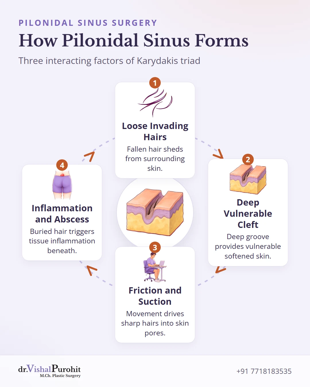
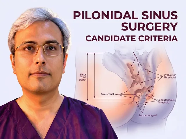
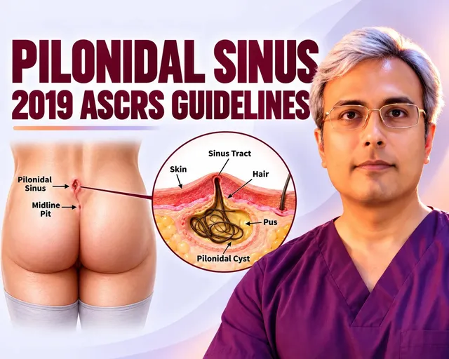
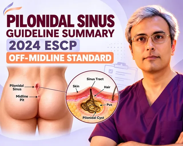
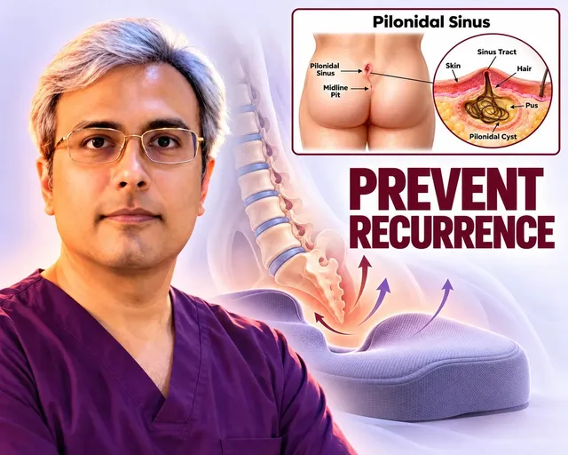
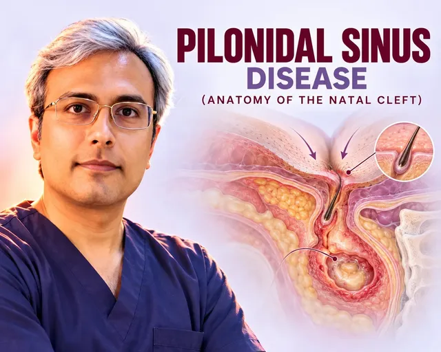
Pilonidal sinus disease is an acquired inflammatory condition of the natal cleft, the deep groove between the buttocks above the tailbone. Rather than an inborn defect, it begins when loose hair pierces vulnerable skin, creating fine surface openings and tunnels beneath.
Key points
- Pilonidal sinus disease is an acquired condition where loose hair pierces the skin of the natal cleft.
- Trapped hair nests create tracts and abscesses that require surgical excision rather than antibiotics.
- Onset peaks between ages 15 and 30, driven by cleft anatomy, friction, and shed hair.
In understanding the anatomy of pilonidal sinus disease, surface pits feed into underlying tracks of scar and trapped debris. Antibiotics cannot dissolve these trapped hair nests, which is why surgical excision of the entire tract system is required for chronic disease.
The condition develops through three interacting elements known as the Karydakis triad:
- Loose invading hairs shed from the scalp, back, or surrounding skin.
- A deep intergluteal cleft providing vulnerable, softened skin.
- Buttock friction creating negative suction that drives sharp hair shafts inward.
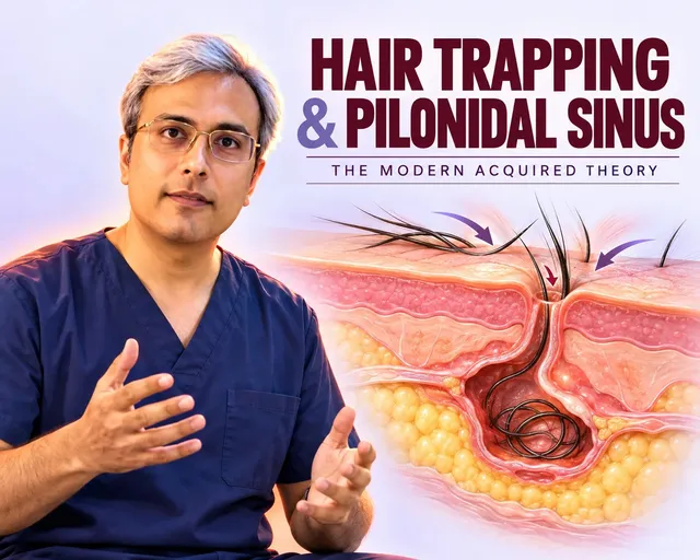
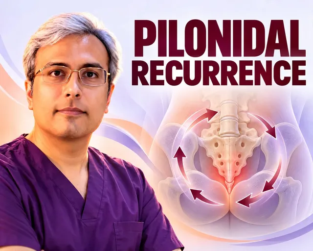
Walking and sitting create a vacuum suction effect that pumps fallen hair root-first into softened pores, showing how hair trapping starts a sinus. Once buried inside fat, the foreign hair causes inflammation and painful abscess formation.
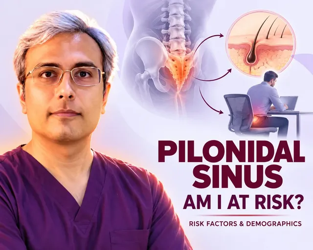
Onset peaks between ages 15 and 30, with reports showing two to three men affected for every woman. These known disease risk factors align with daily habits and anatomy, often aggravated by deep clefts, coarse hair, extra weight, and long seated hours.
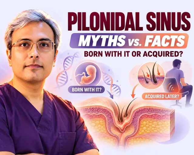
Many misconceptions surround this diagnosis, but pilonidal myths versus facts show that tracts lack hair roots and are neither contagious nor caused by poor hygiene alone. Dropping guilt helps families seek prompt surgical mapping before sinus tunnels branch deeper into tissue.
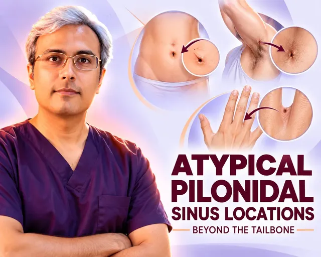
Shed hair can also lodge into other frictional body folds. In atypical pilonidal sinus locations, rare presentations occur in the navel, armpits, groin, and barbers' finger webs. An ultrasound separates these hair-bearing tracts from other conditions such as urachal remnants or cysts before treatment.
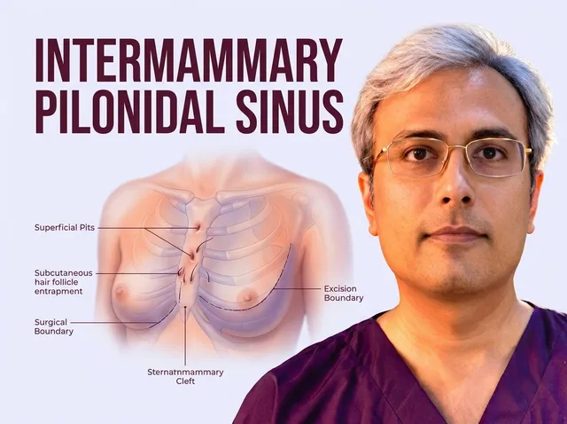
One rare non-sacrococcygeal presentation is intermammary pilonidal sinus disease, which occurs in the crease between the breasts. Opposing skin friction and tension trap shed hair, requiring careful excision and tension-free plastic surgical closure to restore smooth contours.
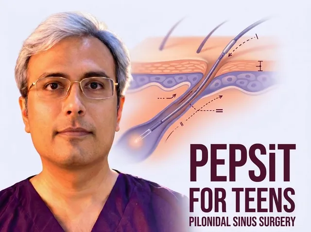
Because onset peaks in young people, parents frequently consider whether pilonidal surgery is safe for teenagers. Rapid growth and seated study raise risks, and pediatric literature describes techniques like PEPSiT as well as flap repairs tailored to tissue maturity to keep recurrence low.
For extensive or recurring conditions, reconstructive flap procedures eliminate the problem. In Jaipur, Dr. Vishal Purohit performs Limberg and Karydakis flap surgery to clear infected tracts, obliterate dead space, and shift scars off the midline.
Symptoms, Diagnosis and When It Becomes Urgent
Pilonidal sinus disease begins quietly in the buttock cleft. Recognizing how tissues change helps patients seek timely evaluation before severe infections take hold.
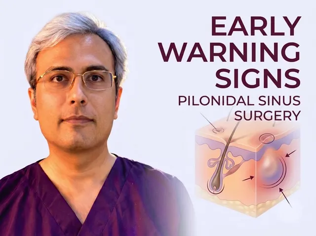
Early changes frequently appear as tiny skin indentations or pits that may not hurt at first near the base of the spine. Recognizing these subtle early symptoms of pilonidal sinus disease helps identify problems before complications arise.
Is this you? You may notice these initial signs:
- Serosanguinous discharge staining undergarments.
- Tenderness while sitting for extended periods.
- Local erythema, which is skin redness caused by localized irritation.
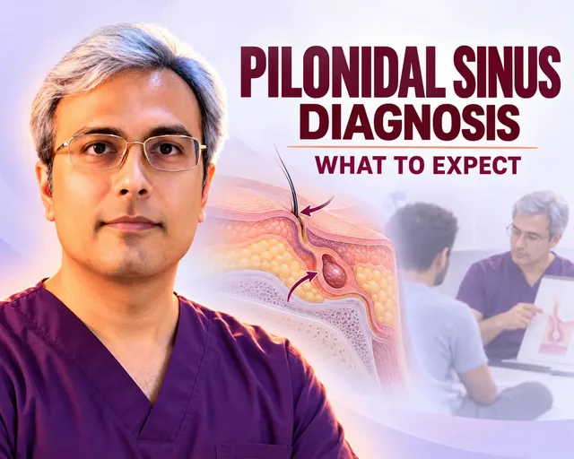
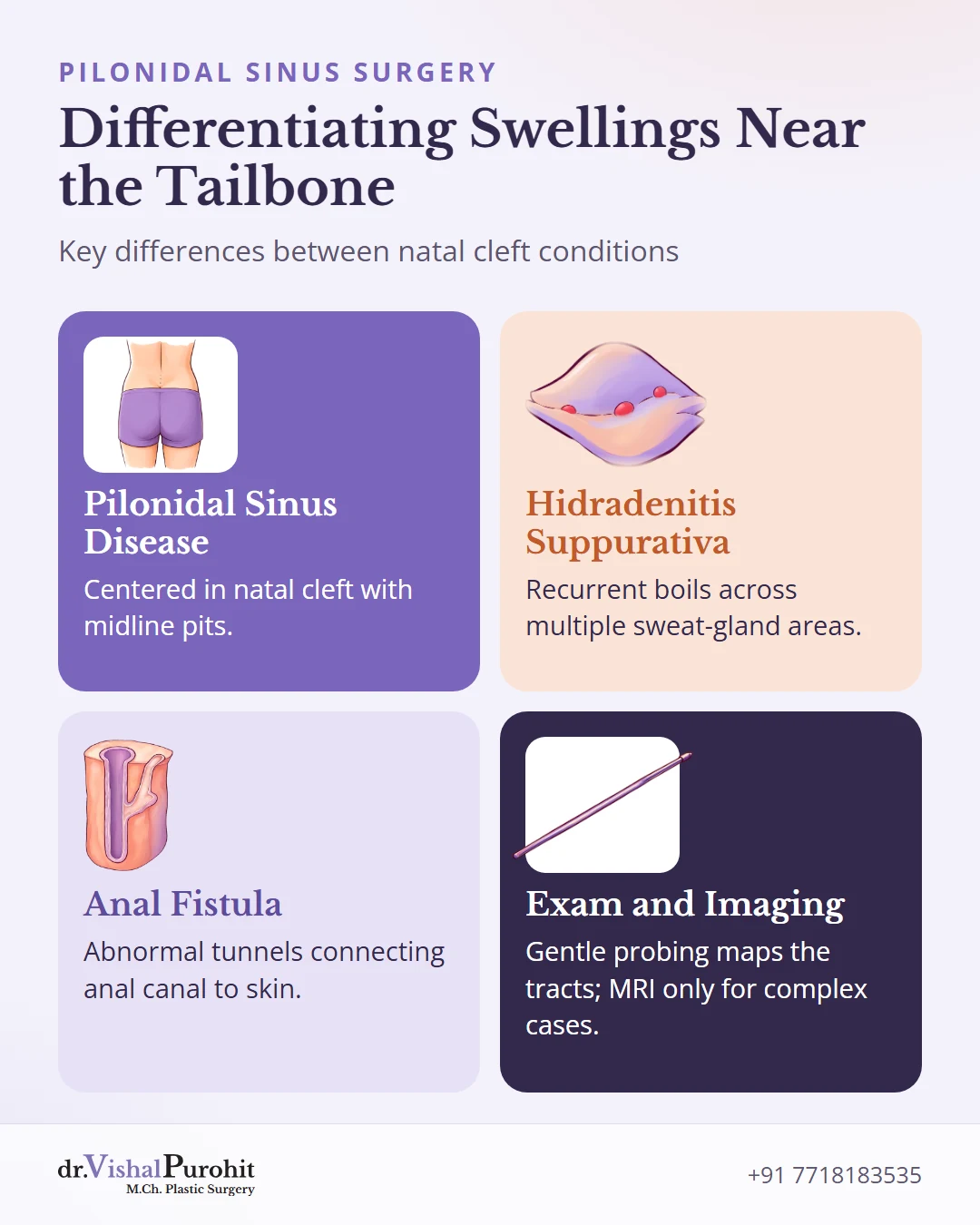
A physical examination confirms the condition by identifying midline pits and feeling subcutaneous cords. During an initial clinical visit, gentle diagnostic probing traces tract depth and side branches, while imaging like MRI is reserved for complex presentations.
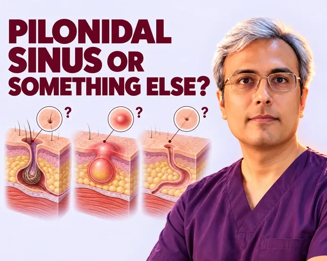
Not every painful swelling near the tailbone originates from pilonidal disease. Other conditions share similar symptoms, requiring careful differentiation:
- Pilonidal sinus disease remains centered inside the natal cleft with midline pits.
- Hidradenitis suppurativa causes recurrent boils across multiple sweat-gland areas, such as the armpits or groin folds.
- Anal fistulas create abnormal tunnels connecting the anal canal to the skin near the anal verge.
Before planning cleft surgery, distinguishing pilonidal disease from other conditions ensures non-cleft issues receive appropriate colorectal or dermatological referrals.
Call the surgical team immediately if you develop an acute abscess with throbbing pain, localized heat, swelling, or active purulent infection.
When bacteria become trapped beneath the skin, an acute abscess can develop rapidly with throbbing pain, localized heat, and swelling. Seeking same-day incision and drainage under local anaesthesia provides immediate pressure relief through an off-midline opening in a brief fifteen to thirty minute procedure.
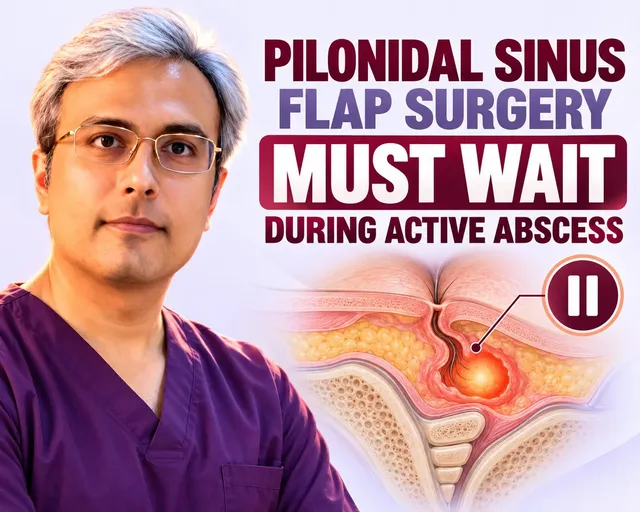
Major reconstructive surgery cannot take place during active purulent infection because inflamed tissue holds sutures poorly. Clinical guidelines recommend clearing acute inflammation first and delaying flap repair until local tissues become soft and calm.
Many patients wonder whether conservative measures can resolve the problem entirely. While improved hygiene and hair control temporarily manage flare-ups, a pilonidal sinus cannot heal permanently without surgery because non-operative care cannot eliminate embedded hair nests or fibrous tracts.
Surgical planning depends on sinus complexity, tissue quality, and prior interventions. Evaluating these personal factors determines whether an individual is a candidate for definitive reconstructive flap techniques when experiencing recurrent drainage, severe discomfort, or multiple flare-ups.
Why Flap Surgery Is Preferred for Pilonidal Sinus
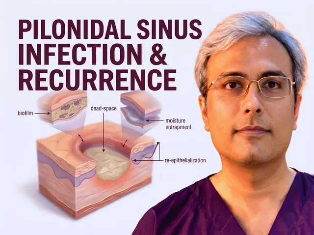
Pilonidal sinus disease is an acquired inflammatory condition in the sacrococcygeal natal cleft, driven by hair penetration, friction, and maceration (softening of wet skin). Midline cuts often fail because stitches sit under constant pull inside a warm, moist fold.
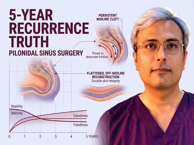
Traditional procedures remove diseased tissue but leave the deep cleft intact. However, leaving the midline groove unchanged allows loose hairs to collect and pierce soft healing skin once again.
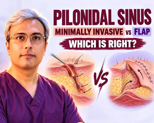
Reconstructive flaps solve this mechanical flaw by shifting well-vascularized healthy skin across the void. This technique flattens the deep valley, fills dead space, and moves the scar off-centre to relieve tension.
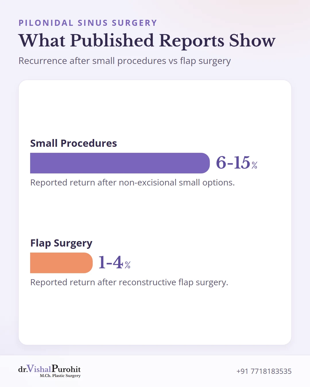
Many patients initially consider small procedures to avoid longer initial recovery. When evaluating small procedures versus flap surgery, published reports note return rates around 6 to 15% for non-excisional small options compared to 1 to 4% for reconstructive flap surgery.
These durability differences become clear over time. Tracking patients over a full five-year follow-up reveals late issues that short-term twelve-month reports miss.
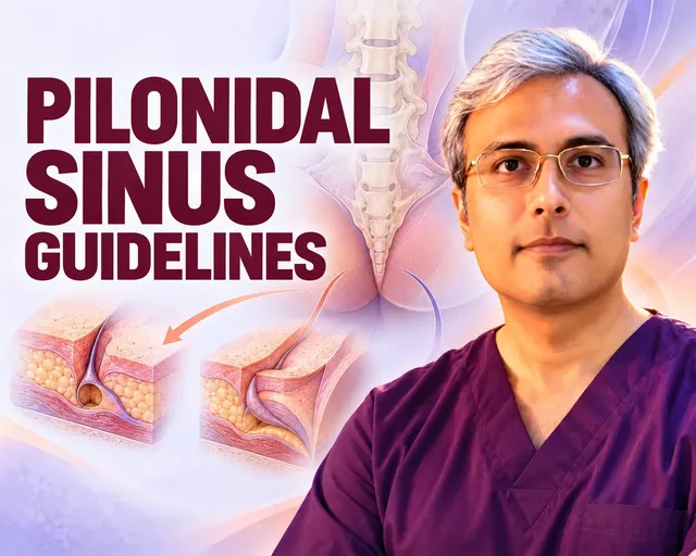
Major international guidelines support off-midline reconstruction. The ASCRS 2019 clinical practice guideline provides strong recommendations for off-midline flap closure over midline closure in complex or repeat cases.
European experts reached a matching conclusion after screening 1025 studies. The ESCP 2024 guideline confirms that off-midline closure is preferred for definitive treatment, while direct midline stitches should be avoided.
When comparing American and European pilonidal guidelines, both panels agree on the foundational principles: prompt off-midline abscess drainage first and reconstructive off-midline flaps for advanced or recurrent disease.
Advanced Surgical Flap Options
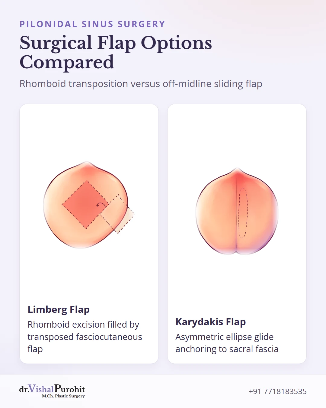
Reconstructive flap surgery clears diseased tissue and replaces it with well-vascularized healthy skin and fat. By flattening the deep natal cleft and moving the final stitch line off the midline, these procedures eliminate the moist friction zone where loose hairs collect and cause recurrent infection.
Limberg and Modified Limberg Flaps
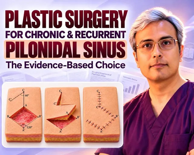
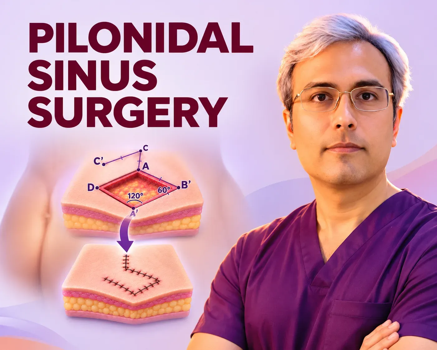
- Excises sinus tracts inside a 60-degree and 120-degree rhomboid diamond
- Swings a fasciocutaneous flap across the gap to fill dead space tension-free
- Modified design shifts the lower tip 1 to 2 cm off-midline away from perianal dampness
- Proven transposition design for durable obliteration of deep, recurrent cleft defects
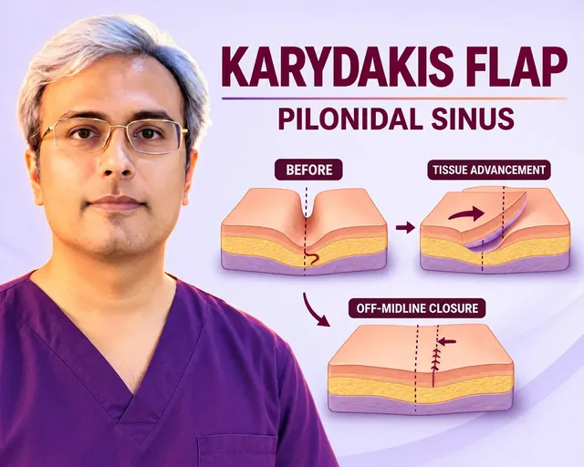
Karydakis Off-Midline Flap
- Excises an uneven ellipse containing midline pits and tracts down to presacral fascia
- Undermines a full-thickness adipocutaneous flap of skin and deep fat on the healthy side
- Glides flap across the midline and anchors directly to sacral fascia with deep stitches
- Places the healed line onto the calmer buttock slope with layered subcuticular closure
Limberg and Modified Limberg Flaps
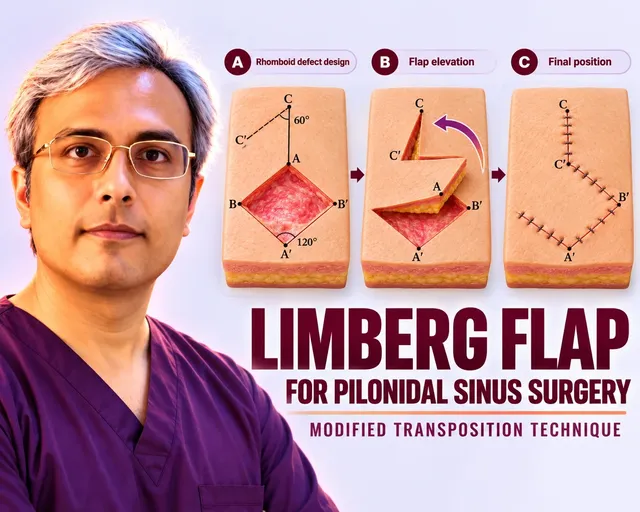
The Limberg flap removes the sinus tracts inside a 60-degree and 120-degree rhomboid diamond. The surgeon raises a fasciocutaneous flap, including gluteal fascia, and swings it across the gap to achieve a tension-free closure that completely fills dead space.
In our clinic, the modified Limberg flap design shifts the lower tip 1 to 2 cm to one side of the midline. This critical adjustment keeps the lowest point of the scar away from damp perianal skin, reducing moisture softening and protecting early healing.
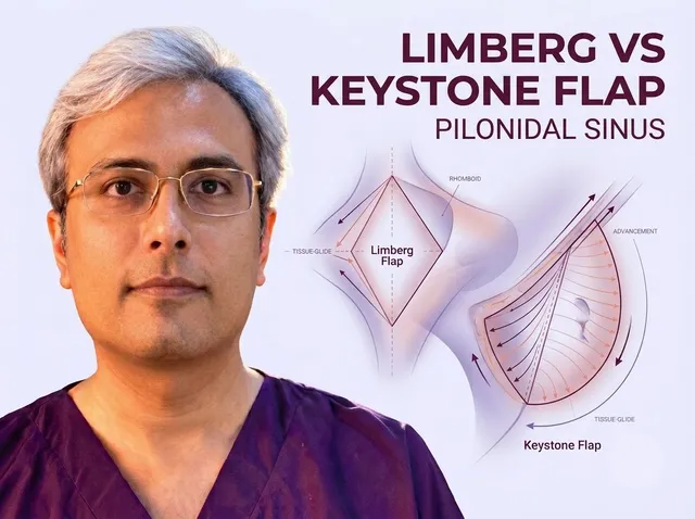
Published literature reviews the Limberg flap vs Keystone flap comparative recovery, noting that while Keystone advancement is described in studies, the Limberg transposition flap remains proven for durable obliteration of deep, recurrent cleft defects.
Karydakis Off-Midline Flap
The Karydakis flap excises an uneven ellipse containing all midline pits, tracts, and diseased tissue down to the presacral fascia over the tailbone. A full-thickness adipocutaneous flap of skin and deep fat is then undermined on the healthy side.
The mobilized flap glides across the midline and anchors directly to the sacral fascia with deep dissolving stitches. Learning how the Karydakis flap moves the scar shows how this flat slide keeps tension minimal and places the healed line onto the calmer buttock slope.
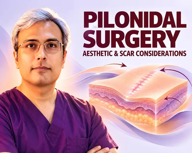
Careful surgical planning and layered subcuticular suturing play a decisive role in protecting cosmetic outcomes and scar appearance, preserving natural gluteal contour while avoiding surface railroad marks.
Other Techniques You May Read About
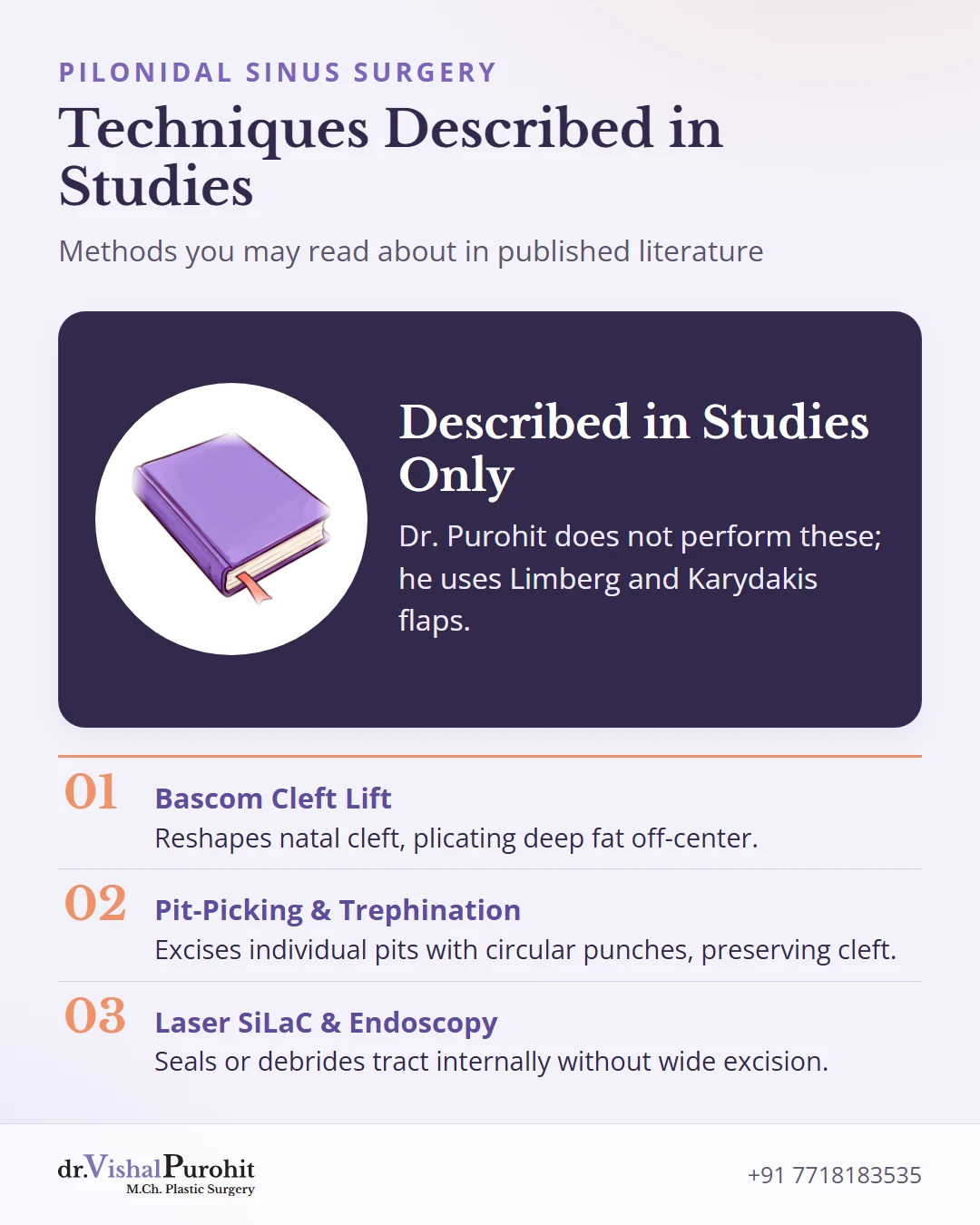
While researching pilonidal disease, you may encounter several alternative procedures described in medical studies. Dr. Vishal Purohit does not perform these techniques at his Jaipur practice.
Key points
- Published medical studies describe several alternative surgical and minimally invasive techniques for pilonidal disease.
- Dr. Vishal Purohit does not perform these alternative procedures at his Jaipur practice, focusing instead on Limberg and Karydakis off-midline flaps and emergency drainage.
- These summaries offer neutral educational context regarding how different flaps, laser, endoscopic, and chemical methods approach the condition.
Instead, he focuses exclusively on proven Limberg and Karydakis off-midline flaps, alongside emergency drainage for acute abscesses. The following summaries provide neutral educational context from published reports.
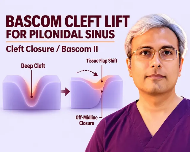
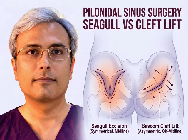
The Bascom cleft lift procedure reshapes the natal cleft by removing diseased skin, plicating deep fat to raise the groove floor, and closing healthy skin off the center. Literature notes high success rates in specialized centers, but it requires specific anatomical trade-offs.
Some reports compare different geometric cut patterns, such as the curved incision in a seagull excision vs cleft lift study. While both position the scar away from the cleft floor, they alter natal cleft depth to varying degrees.
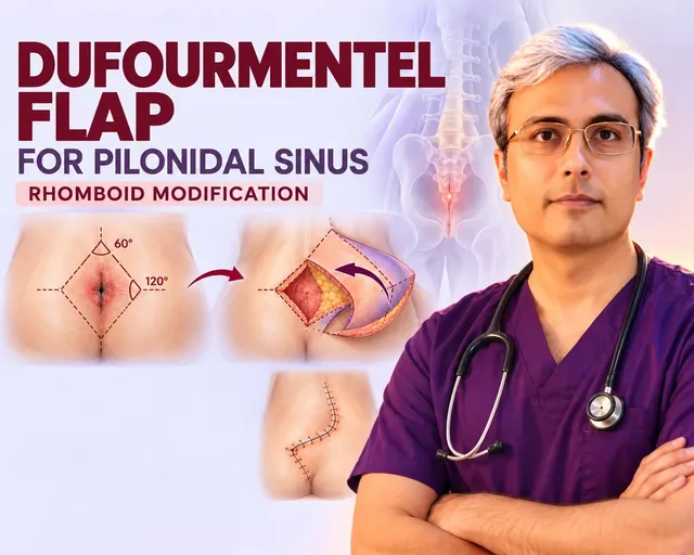
The Dufourmentel flap pilonidal reconstruction modifies the classic rhomboid design by altering internal angles. Published series describe using its broad base and variable geometry to fill wide, stiff, or unusually shaped surgical gaps after extensive tissue clearance.
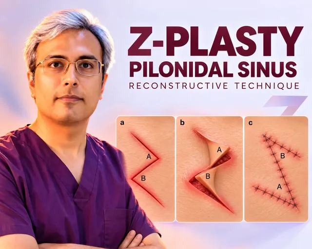
Another plastic surgery concept described in literature is the Z-plasty pilonidal sinus surgery, which transposes two triangular skin flaps. This maneuver redirects tension lines, slightly elevates the cleft valley, and angles the scar across the midline for modest primary defects.
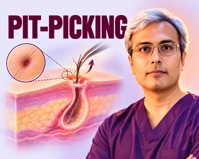
For early-stage disease with minimal branching, studies explore minimal options such as the pit-picking trephination Gips procedure. This office-based method excises individual pits using circular punches and cleans the cavity, though it leaves underlying cleft depth and moisture mechanics completely unaltered.
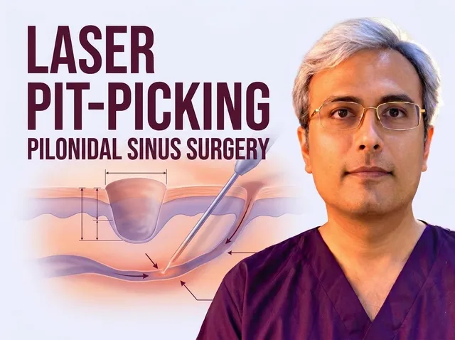
Certain clinical papers examine combining minimal punch excision with thermal energy, described in reports on pit-picking with laser for pilonidal sinus disease. These trials report rapid return to desk work, though deep anatomical hair-trapping forces remain unaddressed.
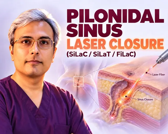
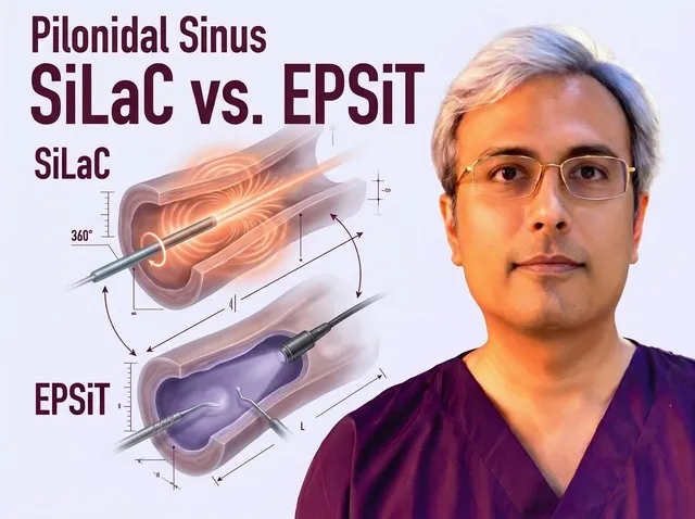
Tract-focused photocoagulation is detailed in literature concerning laser pilonidal sinus closure SiLaC. A flexible radial diode probe delivers circular heat to seal the tract from within, appealing to patients seeking short downtime, despite leaving the intergluteal valley unchanged.
When evaluating non-excisional technologies, comparative reviews such as laser pilonidal sinus surgery SiLaC vs endoscopic EPSiT contrast radial laser thermal sealing with video-guided endoscopic debridement of hidden hair nests and lateral pits.
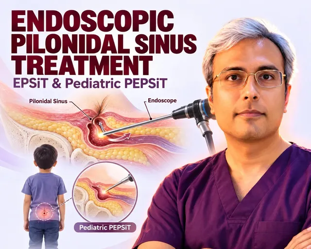
Keyhole visual clearance is also evaluated in studies detailing EPSiT endoscopic pilonidal surgery. Surgeons introduce a video fistuloscope through millimetric ports to directly visualize, extract hair collections, and cauterize the cavity lining without wide skin excision.
Non-surgical chemical techniques outlined in guidelines include phenol and fibrin glue pilonidal treatment. These methods attempt to chemically de-epithelialize tracts using crystal or liquid agents, or seal cleaned spaces with biological glue, often requiring repeat clinic applications.
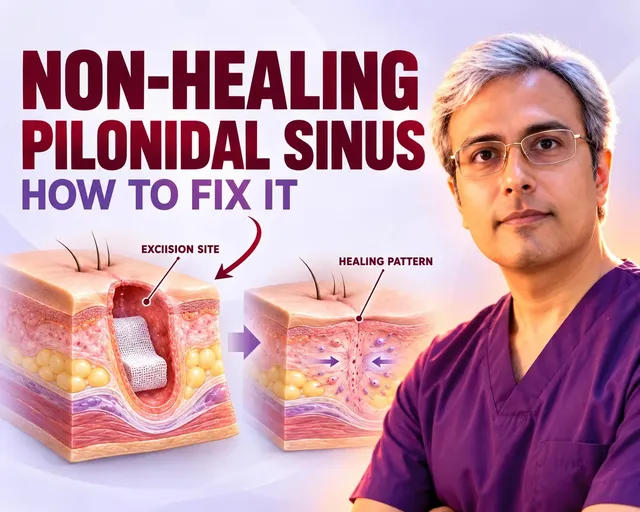
Finally, when previous operations break down or stall in the midline, studies on non-healing pilonidal wound repair emphasize converting chronic granulating defects into durable closures. Converting open, stalled midline wounds relies on vascularized off-midline flaps to flatten the cleft and restore skin coverage.
Preparing for Surgery and the Day of the Operation
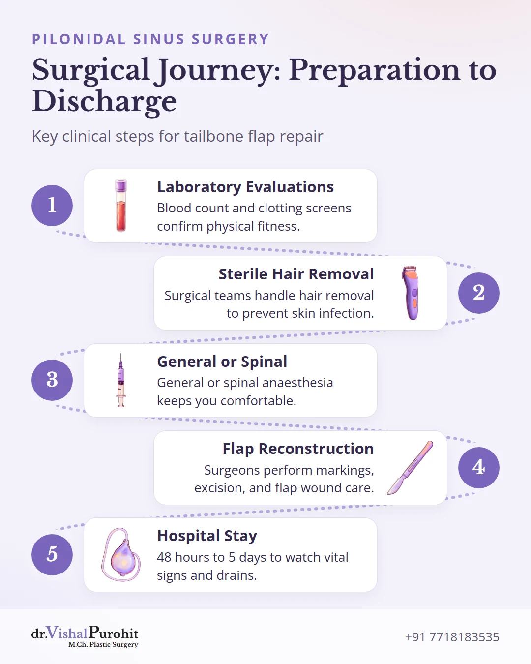
Thorough preparation ensures your tailbone procedure is safe, organized, and focused on smooth healing. Clear clinical routines protect your skin, confirm internal fitness, and guide your surgical steps from admission to discharge.
Key points
- Careful pre-operative hygiene, professional hair removal, and laboratory health checks protect healing and ensure surgical fitness.
- Targeted anaesthesia and nerve blocks manage pain during the procedure, which is followed by a hospital stay of 48 hours to 5 days to monitor drains and vitals.
- Clear financial planning provides itemized estimates to streamline insurance approvals and prevent surprises.
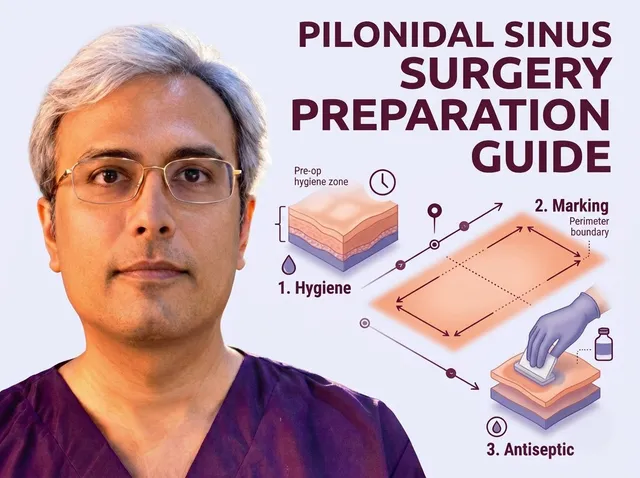
At home, careful skin care reduces bacteria before your procedure. Follow clinical guidance on hygiene, hair removal, and medicines to protect fragile skin and follow fasting hours.
Do not shave the tailbone area yourself, as razors create small nicks that invite infection. Instead, surgical teams handle hair removal with sterile tools.
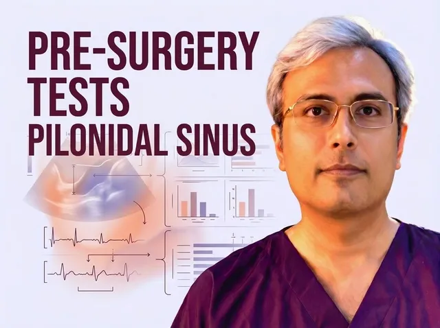
Before any surgical booking, laboratory evaluations assess your general physical stability. Essential medical tests and health checks, such as a complete blood count and clotting screens, confirm that anemia or hidden infections are managed early. If an acute abscess is present, emergency drainage must take place before definitive flap repair.
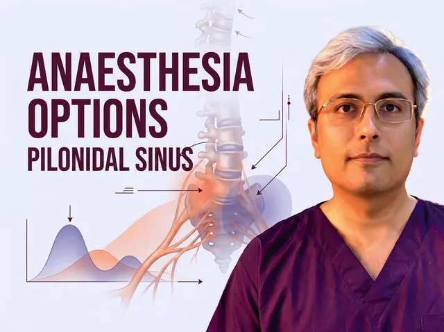
During the operation, Dr. Vishal Purohit uses general or spinal anaesthesia to keep you comfortable. Spinal anaesthesia numbs the lower body while you stay awake and pain-controlled. Studies also describe local, spinal, and sacral ESP block options for pain relief.
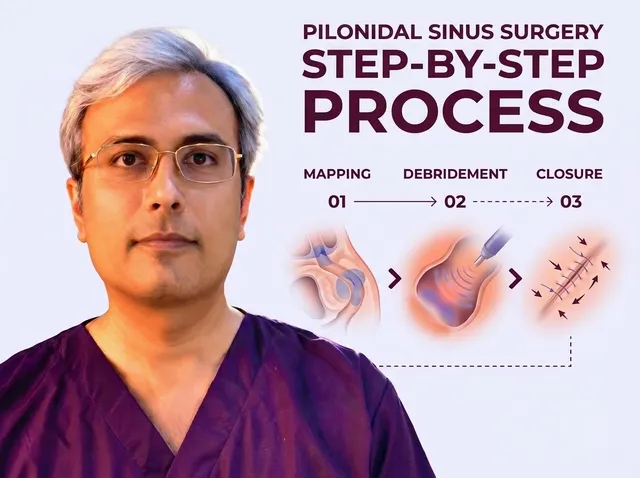
Understanding each hospital milestone helps you feel confident on the day of treatment. Your care pathway follows key stages from anaesthesia to discharge:
- Geometric markings precise flap boundaries
- Anaesthesia targeted nerve blocks
- Excision sinus tract removal
- Wound care closed-suction drain placement
- Discharge 48 hours to 5 days in hospital
Flap reconstruction patients stay in hospital for 48 hours to 5 days to monitor vital signs and manage a closed-suction drain before meeting safe discharge criteria.
Finally, clear financial planning prevents surprises before your admission date. Evaluating pilonidal surgery costs in Jaipur helps you compare item-wise estimates covering operating theatre time, anaesthesia, hospital stay, and dressings.
Having a written breakdown also streamlines medical insurance approvals and ensures continuous post-operative follow-up support.
Recovery & Healing Timeline
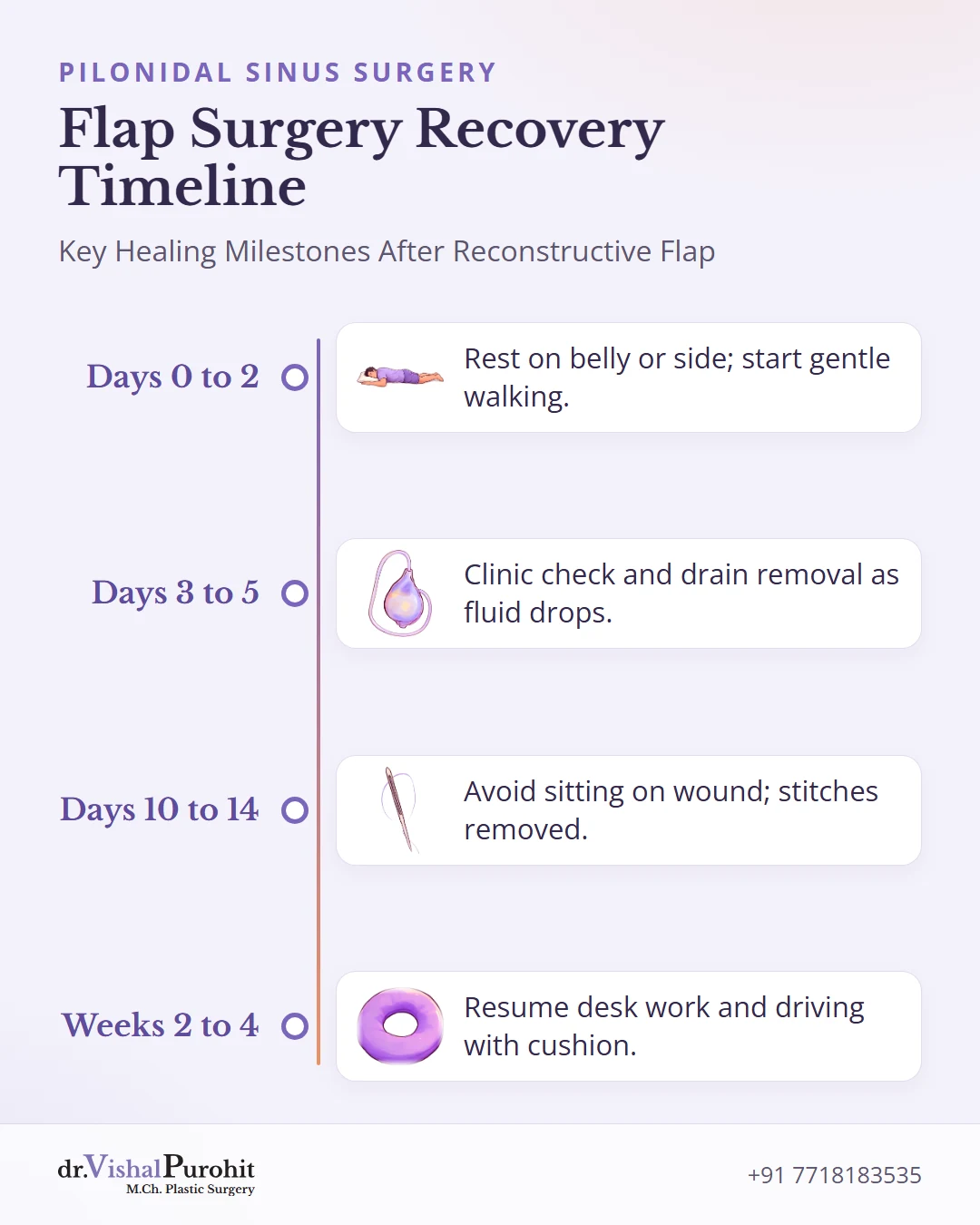
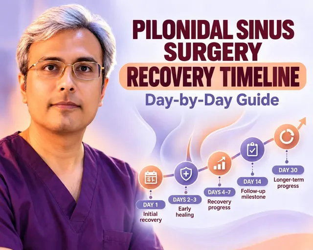
Reconstructive flap surgery closes the wound primarily instead of leaving an open space to fill over months. Healing follows steady steps under close surgical guidance.
A closed-suction tube stays beneath the flap to draw out pooling blood and fluid. Daily stripping, flat bulb suction, and measuring fluid output are part of standard flap drain care at home until clinic removal.
Daily routines return in clear stages as your tissues integrate:
- Days 0 to 2 Rest on belly or side; gentle indoor walking
- Days 3 to 5 Clinic check and drain removal once output drops
- Days 10 to 14 Avoid sitting directly; stitches removed days 12 to 14
- Weeks 2 to 4 Graded desk work and driving resume with cushion
Following this progressive surgery recovery timeline helps protect the healing flap edges while sitting stamina and activity steadily improve.
While primary flap wounds heal directly, stalled repairs or wide spaces may utilize advanced pilonidal wound healing techniques such as vacuum-assisted closure (VAC) dressings.
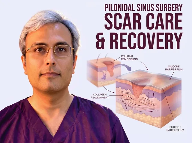
As the off-midline cut seals, understanding how flap incisions heal over time helps guide sun cover and loose clothing to soften the maturing scar line.
Risks, Warning Signs and Preventing Recurrence
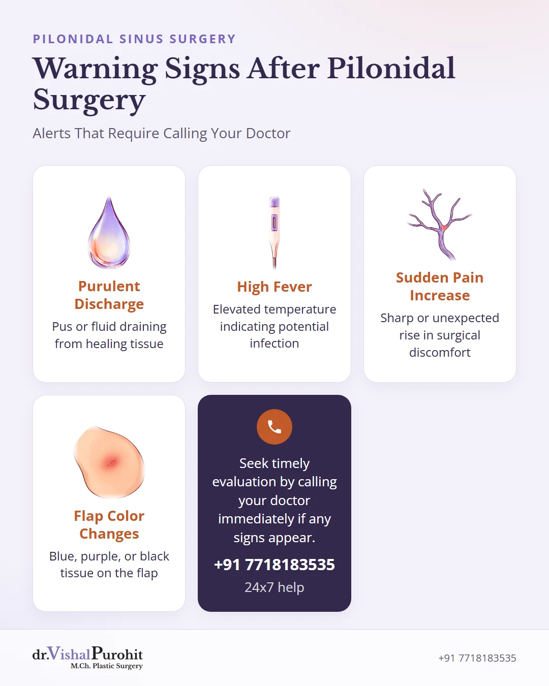
Understanding potential surgical complications, recovery alerts, and protective habits helps you heal with confidence after pilonidal sinus treatment. Careful aftercare and reconstructive planning lower your chances of setbacks.
Key points
- Being aware of complications helps you spot risks like infection, fluid collections, or wound opening early.
- Certain warning signs require prompt medical contact to protect healing tissue.
- Protective lifestyle habits help prevent recurrence, and studies describe laser hair removal after healing.
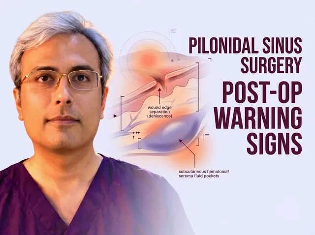
Every procedure carries potential concerns in the sacrococcygeal area between the buttocks. Being aware of the risks of pilonidal sinus surgery helps you identify possible complications:
- Surgical site infections
- Fluid collections
- Wound dehiscence, where skin edges split open
Call the surgical team immediately if you notice purulent discharge, a high fever, sudden increases in pain, or color changes such as blue, purple, or black tissue on a flap.
Recognizing early problems protects healing tissue. Contact your doctor promptly if you notice these warning signs after surgery:
- Purulent discharge
- A high fever
- Sudden increases in pain
- Color changes such as blue, purple, or black tissue on a flap
Daily habits shield the surgical result once your incision closes. Protective measures to prevent pilonidal sinus return reduce friction and pressure through simple steps:
- Taking regular standing breaks
- Weight control
- Wearing cotton underwear
- Stopping smoking before and after flap surgery
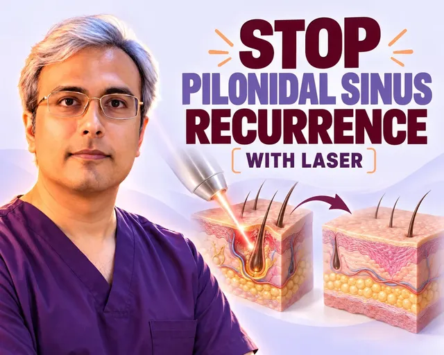
Managing hair entry addresses the primary cause of recurrent tracts. Undergoing laser hair removal across the cleft and surrounding skin slopes once the wound has fully closed destroys deep hair bulbs so loose shafts cannot pierce delicate skin.