
In short
- The Limberg flap removes disease as a diamond and swings nearby healthy skin across the gap.
- The modified version shifts the lower tip 1 to 2 cm off the midline to protect skin near the anus.
- Dr. Vishal Purohit performs Limberg and Karydakis flap surgery in Jaipur, matched to each patient’s shape.
Who this surgery is for
The Limberg flap is an off-midline repair for pilonidal sinus disease. Off-midline means the final scar rests away from the centre line. It suits wide disease, far-side tracts, large defects, and redo cases where prior midline surgery failed.
The problem it solves is mechanical. A deep buttock cleft traps loose hair. Friction drives hair into midline skin. A scar left in that damp groove breaks down and restarts the cycle. The Limberg flap breaks the cycle two ways: it flattens the cleft and it moves the scar onto drier side skin.
This page is for two groups. The first is patients with broad or branching disease advised flap surgery. The second is patients whose open excision or midline closure failed elsewhere and left a wide or low defect. The fold brings its own blood supply, so it covers ground a simple slide cannot.
One fact shapes this whole page. Dr. Vishal Purohit performs Limberg and Karydakis off-midline flap surgery in Jaipur, plus urgent abscess drainage. The clinic does not offer phenol, SiLaC laser, endoscopic or pit-picking methods, Bascom cleft lift, Dufourmentel flap, or Z-plasty. Those appear here only as published background where a comparison is needed.
The classic diamond: exact shape, folded healthy skin
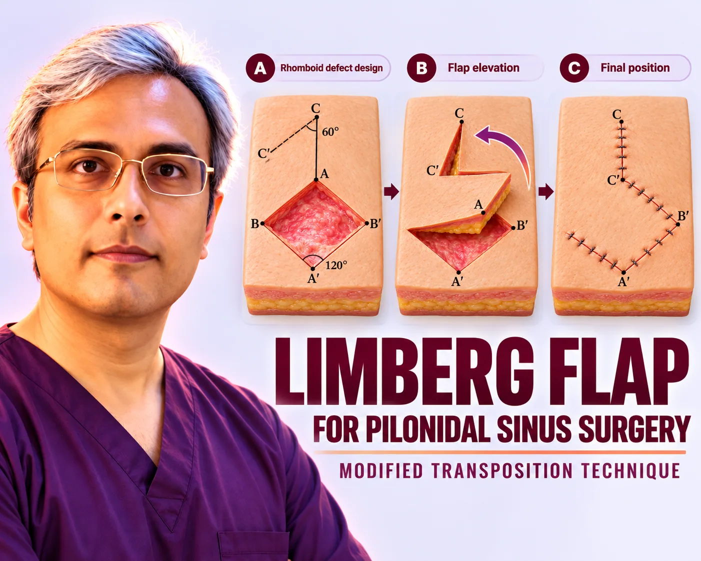
The classic Limberg design starts with a rhomboid, a diamond with 60-degree and 120-degree corners. The surgeon draws it around every pit, tract, and scar, with the long axis along the cleft. Exact angles matter. They let the donor flap fit the defect like a puzzle piece, with no stretch and no leftover gaps.
Next the surgeon raises the flap. This is a fasciocutaneous flap, meaning skin plus the firm gluteal fascia beneath it. Fascia is the tough sheet over muscle. Including it gives the flap strength and a rich blood net. The flap is cut on three sides and hinged on the fourth, then swung across the defect. Transposition is the technical word for this swing.
Because the flap carries its own supply, the folded edge stays pink and heals fast. This is why the Limberg covers wide defects well. A plain side-to-side stitch would pull tight over a big gap. The fold fills the gap with fresh local tissue instead. The donor site, where the flap lifted from, closes directly since the buttock skin there is loose.
The cut goes down to the presacral fascia over the tailbone. All diseased tissue inside the diamond comes out in one piece: tract walls, pus pockets, hair nests, and swollen fat. Bleeding is stopped point by point, because blood trapped under a folded flap blocks sealing.
The modified turn: lower tip 1 to 2 cm to the side
The modified Limberg flap (MLF) changes one thing: the lowest corner of the diamond. In the classic drawing this tip sits on the midline, close to the anus. The modified drawing shifts it 1 to 2 cm sideways. Everything else — the diamond, the fold, the layered closure — stays the same.
Only the lower tip moves because only the lower tip causes trouble. The upper and middle scar sits on the buttock slope, which stays dry. The lowest point faces moisture from below. Shifting just that corner keeps the whole scar on calmer ground without redrawing the full design.
Marking the shift takes seconds but needs a steady eye. The surgeon presses the buttocks together, finds where the cleft floor meets drier slope skin, and parks the tip there. Too small a shift leaves the tip in the damp zone. Too large a shift stretches the fold. The 1 to 2 cm range is the reported sweet spot in published descriptions.
Why this centimetre-level shift matters
Skin near the anus stays moist. Moisture softens scars, a process doctors call maceration. A soft scar itches, cracks, and opens. It also traps hair more easily. Moving the lowest scar point 1 to 2 cm sideways puts it on drier skin that resists all three problems.
Low wounds also face more germs. The anal verge sits nearby. A midline tip at that level collects discharge and heals slowly. An off-midline tip drains cleanly and dries faster. This is why surgeons pay most attention to the bottom corner and less to the upper scar, which rarely causes trouble.
The shift does not weaken the repair. The flap still anchors to the deep fascia. The cleft still flattens. The fold still brings fresh supply. One small sideways move buys drier healing at the exact point where classic designs struggled most.
How the operation runs step by step
Care follows a fixed order. Each step prepares the next:
- Mapping under anaesthesia. Face down, spinal or general anaesthesia on board, the surgeon marks every pit, side opening, tract, and old scar.
- Diamond drawing. The 60 and 120-degree rhomboid is drawn around the full disease field, with the lower tip parked 1 to 2 cm off the midline.
- Excision to fascia. The disease block is removed down to the presacral fascia in one piece, and bleeding is stopped point by point.
- Flap raising. The fasciocutaneous flap, skin plus gluteal fascia, is cut on three sides and lifted on its hinge with supply intact.
- Transposition. The flap swings across the gap and settles with no pull; the donor site edges come together directly.
- Layered closure. Deep stitches anchor the flap to fascia, middle layers close small gaps, and the skin closes with stitches or staples while a thin suction drain exits through a side cut.
Dressing follows a buttock-press check: a shallow cleft with the zigzag scar clearly off centre. The drain stays on gentle suction for the first days. It is standard care, not a sign of trouble. Even a well-built fold sheds early fluid, and removing that fluid speeds sealing.
Results: fast filling and under 3 percent return
Healing runs fast because the closure is tension-free. Tension-free means no pulling at the wound edges. Most patients walk the same day. The drain leaves in the first week. Desk work returns in 2 to 3 weeks. Full sport waits 6 to 8 weeks until wound review allows it.
Published series report return rates below 3% after Limberg flap surgery (reported endpoint as published). The flat cleft and side-lying scar explain the figure: hair finds no deep damp groove to re-enter. Small lower-edge slips and fluid pockets are the most reported nuisances, and they usually settle with dressings at review.
Patients often ask how this compares with the Karydakis flap. Pooled trial data show similar return rates for both flaps. The Limberg fold suits wide or far-side defects. The Karydakis slide suits midline-near disease. After healing, laser hair removal after healing guards both results. The full reconstruction hub lists every flap option in one place.
Key figures from published series
- Return rates below 3% are reported after Limberg flap surgery in published pilonidal series (reported endpoint as published).
- The classic design uses a rhomboid with 60-degree and 120-degree corners, closed by fasciocutaneous transposition including gluteal fascia.
- The modified version parks the lower tip 1 to 2 cm off the midline to keep the lowest scar point off damp perianal skin.
What Dr. Vishal Purohit does in Jaipur
Dr. Vishal Purohit is an M.Ch plastic surgeon treating pilonidal sinus disease in Jaipur, performing definitive Limberg and Karydakis off-midline flap surgery. No phenol, laser, or endoscopic shortcuts; matched to your anatomy.
At consult the surgeon maps pits, tracts, side branches, and old scars. He explains whether the Limberg fold or the Karydakis slide fits the cleft depth and disease spread. Every patient gets a written plan for anaesthesia, drain care, dressing dates, and hair-care teaching. Acute abscesses are drained urgently first, with staged flap repair once swelling settles.
Outstation patients get clear dates: mapping at the consult visit, day-care or short-stay surgery, drain removal in the first week, and reviews at 1, 3 and 6 weeks. Redo and low-perianal cases get longer watch. The flap is chosen for the build seen, never from a fixed menu. This is educational information, not medical advice.
Next step
Patients with wide disease, low tracts, or a failed operation elsewhere can book consultation with Dr. Vishal Purohit in Jaipur for disease mapping with a build-matched Limberg or Karydakis plan. Bring old operation notes and scans where available.