
In short
- Pilonidal sinus is trapped hair and skin debris in the tailbone groove — not a birth defect.
- Midline pits feed tunnels below. Each cycle of blockage brings an abscess and adds scar.
- Treatment means removing all pits and tracts — antibiotics alone cannot do this.
A common disease in a hidden fold
Pilonidal means a nest of hair. The disease lives in the natal cleft — the deep groove between the buttocks above the tailbone. It strikes teens and young adults, most often males aged 15 to 30.
Early signs are often mild. Sticky or bloody discharge from tiny pits. A bad smell. Pain on sitting. Patients are asked to bring old scans and prescriptions, because records of past abscesses show how far the tunnels have spread. Many patients have had one or more burst abscesses before a true sinus ever shows. Grasping this base makes each next choice easier — from how hair trapping starts to why the scar must sit off the midline.
The groove that traps hair
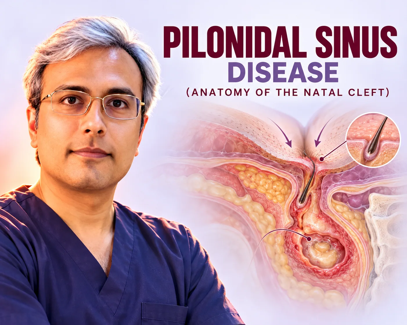
The tailbone area stacks three risk layers. Bone sits at the bottom. Fat and fascia lie above it. Folded skin tops it off in a deep, warm, moist groove. Each step and each sitting hour rubs the two sides together.
In prone people the groove runs deep and narrow. Air flow stays low. Sweat softens the midline skin. Loose hairs — often shed from the head or back — collect in the fold. Then buttock motion drives the sharp hair ends into the softened pores.
Factors that deepen the trap:
- Coarse hair and heavy sweating — common in young males.
- Long sitting hours — study, driving, desk work.
- Extra weight, which deepens the fold and raises moisture.
This is why the old birth-defect idea was dropped. The disease forms; it is not born. The cleft builds the trap. Hair supplies the foreign body. Friction supplies the force. Each treatment, from hygiene to flap surgery, breaks one of these links.
From tiny pits to tunnels
The first visible marks are midline pits. They look like small dark pores in a row of one to several. Each pit marks where a hair pierced the skin and the spot filled in. Pits may ooze lightly for months. Many people ignore them until the first abscess.
Below the pits hides the buried part. It is a network of pockets or branched tunnels in the cleft fat. The tunnels hold nests of hair, debris, and swollen tissue. When pressure builds, the pocket turns into a hot, painful abscess beside the midline.
Two mouth types matter:
- Primary pits — midline entry points, the root of the disease.
- Secondary openings — off-centre exits, where old tracts drain pus.
The natural course runs in cycles. Pits take in hair. The pocket fills. Bacteria form an abscess. Drainage — on its own or by cut — eases pain but leaves pits and tracts behind. Each cycle lays more scar and spreads the tunnels. Early cycles may hold one pit and a pocket under 2 cm. Late cycles can span several centimetres with many exits. So guideline care stages treatment: calm the active infection with acute abscess treatment first, then remove the whole pit-tract system by planned surgery before the map grows.
See a surgeon promptly for these signs
- Repeat discharge, smell, or stained clothes
- Hot swelling that blocks sitting or sleep
- A past surgery wound that never fully closed
Not a true cyst
Under the microscope this is no closed cyst sac. It is a long battle against trapped hair. Tissues show swollen repair zones with giant cells around hair bits. Tract walls carry patchy lining, not a full cyst wall.
This solves three puzzles:
- Why drugs alone fail — antibiotics kill bacteria but leave the hair nest, so oozing returns.
- Why tracts feel woody — the firmness is layered scar, not plain swelling.
- Why left-behind bits bring return — each spared hair nest restarts the reaction.
Treatment must remove the whole system by surgery. Then flap methods fix the trap itself. Excision clears the blockage down to firm fascia. Limberg or Karydakis rebuilding flattens the cleft and shifts the scar onto dry side skin. Small chemical or heat methods reported in journals aim to dry or clean tracts without changing the cleft. They appear here as literature context only. This is educational information, not medical advice.
Key figures from published series
- Pilonidal sinus is a commonly seen disease of young adult males, treated by surgery with growing use of small interventions.
- Limberg and Karydakis rebuilding are the two most used off-midline closure methods for pilonidal disease (systematic review and meta-analysis).
- One phenol study of 80 patients reports success-related rates of 90 to 95% with a 37.5% re-treatment-related figure (endpoint as published) — cited as literature context only; the clinic does not perform phenol.
What Dr. Vishal Purohit does in Jaipur
Dr. Vishal Purohit treats pilonidal sinus disease in Jaipur. Each visit maps cleft depth, pit count, and tract spread against the midline and anal verge. Proven cleft disease is treated with Limberg or Karydakis flap surgery, and hot abscesses are drained promptly.
Signs pointing to other diseases go to the right specialty. The clinic does not perform phenol, laser, endoscopic, pit-picking, Bascom, Dufourmentel, or Z-plasty methods. This is educational information, not medical advice.