
Chest fullness in men usually stems from enlarged breast gland tissue, accumulated chest fat, or a combination of both. Because excess fat feels soft while glandular tissue forms a firm disc behind the areola, physical examination provides the initial clue to what lies beneath the surface.
Understanding the underlying tissue is essential because firm glandular parenchyma does not shrink with exercise alone. Exploring how gynecomastia is diagnosed shows how clinical palpation and imaging distinguish glandular proliferation from fatty fullness to guide appropriate treatment.
Two Conditions That Look Alike
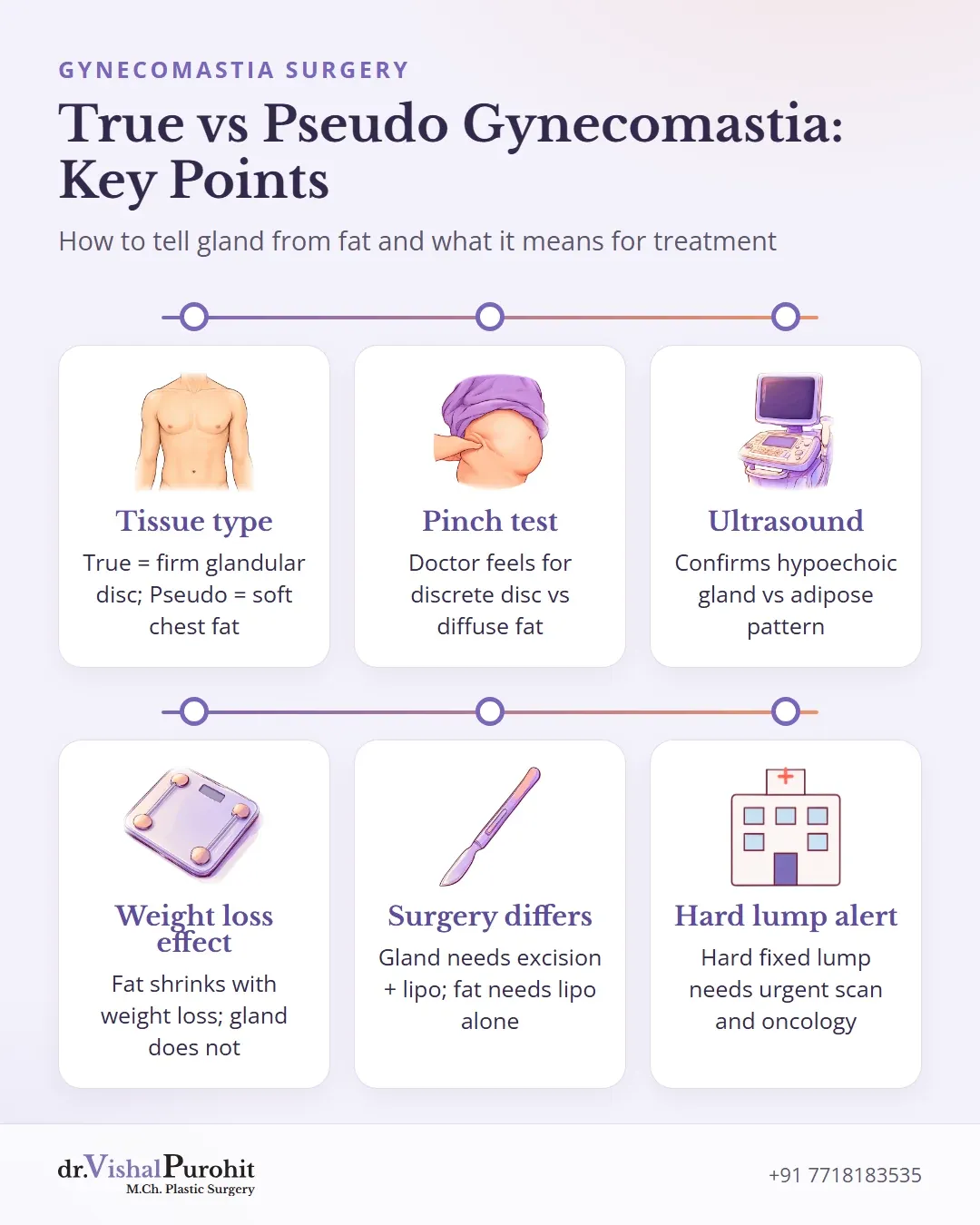
Enlargement of the male chest usually comes down to two different conditions that can look very similar from the outside.
Gland, Fat, or Both
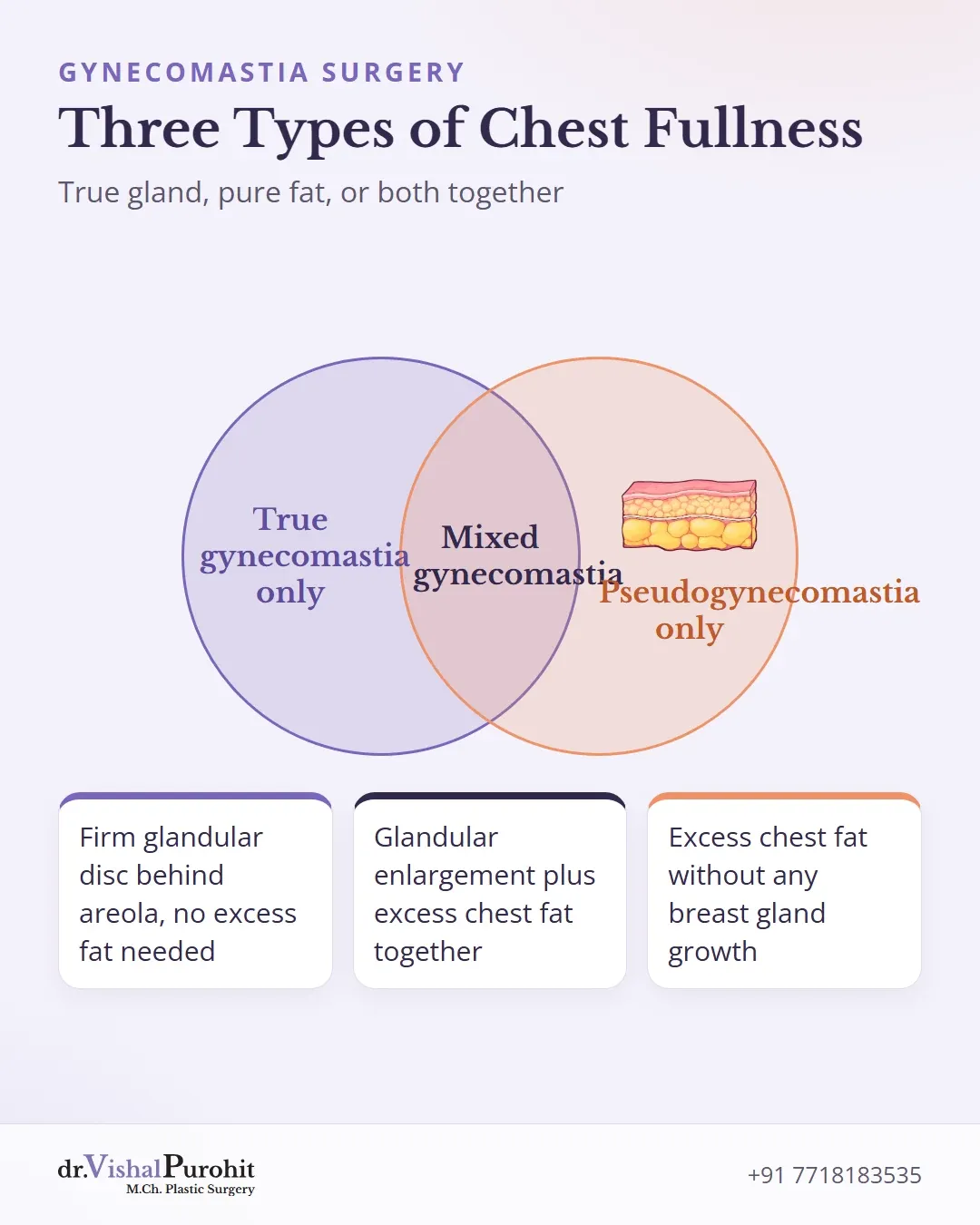
The underlying tissue causes the difference:
True Gynecomastia
This is an abnormal growth of firm, glandular breast tissue, typically forming a palpable disc or button directly behind the areola.
Pseudogynecomastia
This refers to an accumulation of excess chest fat without any growth of breast gland tissue. It accounts for most cases of chest fullness seen in men with higher body weight.
Mixed Gynecomastia
Many men have a combination where glandular enlargement and excess chest fat exist together.
Because fat is soft while breast gland tissue is firm, the two feel noticeably different to the touch. How severity is graded is explained in the guide to gynecomastia grades.
Telling these conditions apart is essential because glandular tissue does not shrink with diet or exercise alone, whereas excess chest fat often responds to weight loss. When surgery is planned, identifying whether gland tissue, fat, or both are present determines the correct treatment approach.
How Each One Feels and Looks
During a physical examination, direct palpation of the chest tissue helps distinguish true glandular enlargement from accumulated chest fat.
Comparison of Key Features
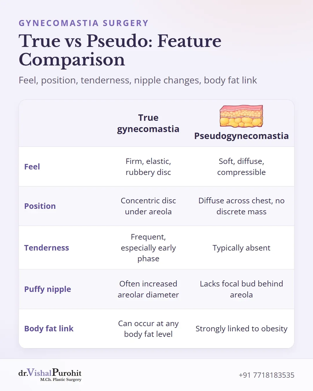
| Feature | True gynecomastia | Pseudogynecomastia |
|---|---|---|
| Feel | Firm, elastic, mobile, rubbery disc | Soft, diffuse, non-nodular, compressible tissue |
| Position | Concentric disc directly beneath the nipple-areolar complex | Diffuse across the chest without a discrete retroareolar mass |
| Tenderness | Frequently present, especially during the early active phase | Typically absent |
| Puffy nipple | Often accompanied by an increased areolar diameter or projection | Lacks a discrete focal bud behind the areola |
| Other body fat | Can occur regardless of body fat, including during puberty | Strongly associated with overall body fat or obesity |
Glandular Tissue Versus Hard Lumps

True gynecomastia presents as a mobile, rubbery or firm disc measuring at least 2 cm beneath the nipple-areolar complex.
Gynecomastia is never a hard lump. A hard, irregular, fixed, or one-sided mass is not typical of benign glandular tissue.
Dr. Purohit’s note: Dr. Purohit’s protocol states that any hard mass suggests conditions such as carcinoma or a tense cyst and requires urgent imaging with ultrasound or MRI, followed by immediate referral to an oncologist.
The Pinch Test and Its Limits
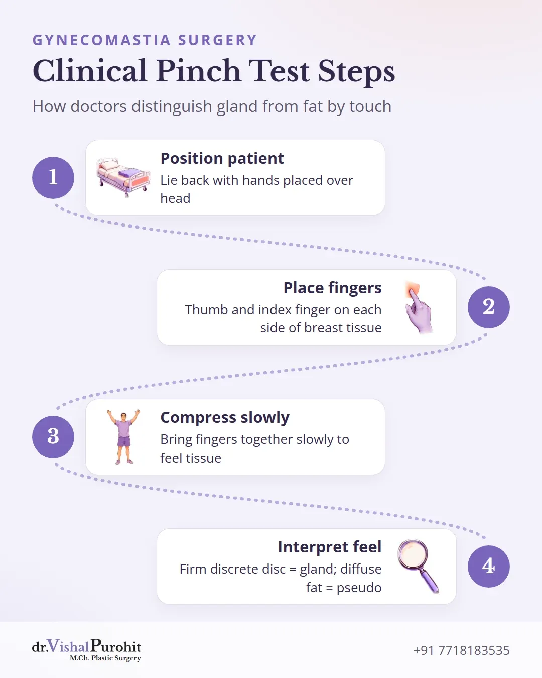
In a clinical evaluation, a physical examination and direct palpation are the first steps to evaluate chest enlargement. During this assessment, a doctor typically has the patient lie back with hands placed over the head. The clinician positions a thumb and index finger on each side of the breast tissue and brings them together slowly.
In true gynecomastia, the examiner feels a firm, discrete, disk-like mound of mobile glandular tissue directly beneath the areola. In contrast, pseudogynecomastia presents with diffuse fatty tissue across the chest without a distinct subareolar nodule.
Why Home Self-Checks Are Inconclusive
Attempting this maneuver at home serves only as a rough gauge and cannot replace a medical evaluation:
- Lack of precision: Subtle glandular enlargement can be difficult for an untrained individual to separate from surrounding adipose tissue.
- Cannot rule out conditions: A home check cannot confirm mixed presentations or exclude other underlying conditions that require clinical palpation or imaging.
When Ultrasound Helps
At his practice in Jaipur, Dr. Vishal Purohit begins the evaluation with a thorough clinical examination and direct palpation of the chest. Ultrasound or MRI is arranged only when imaging is required to clarify the findings, while hormone blood tests are advised strictly if there are other symptoms of hormonal imbalance.
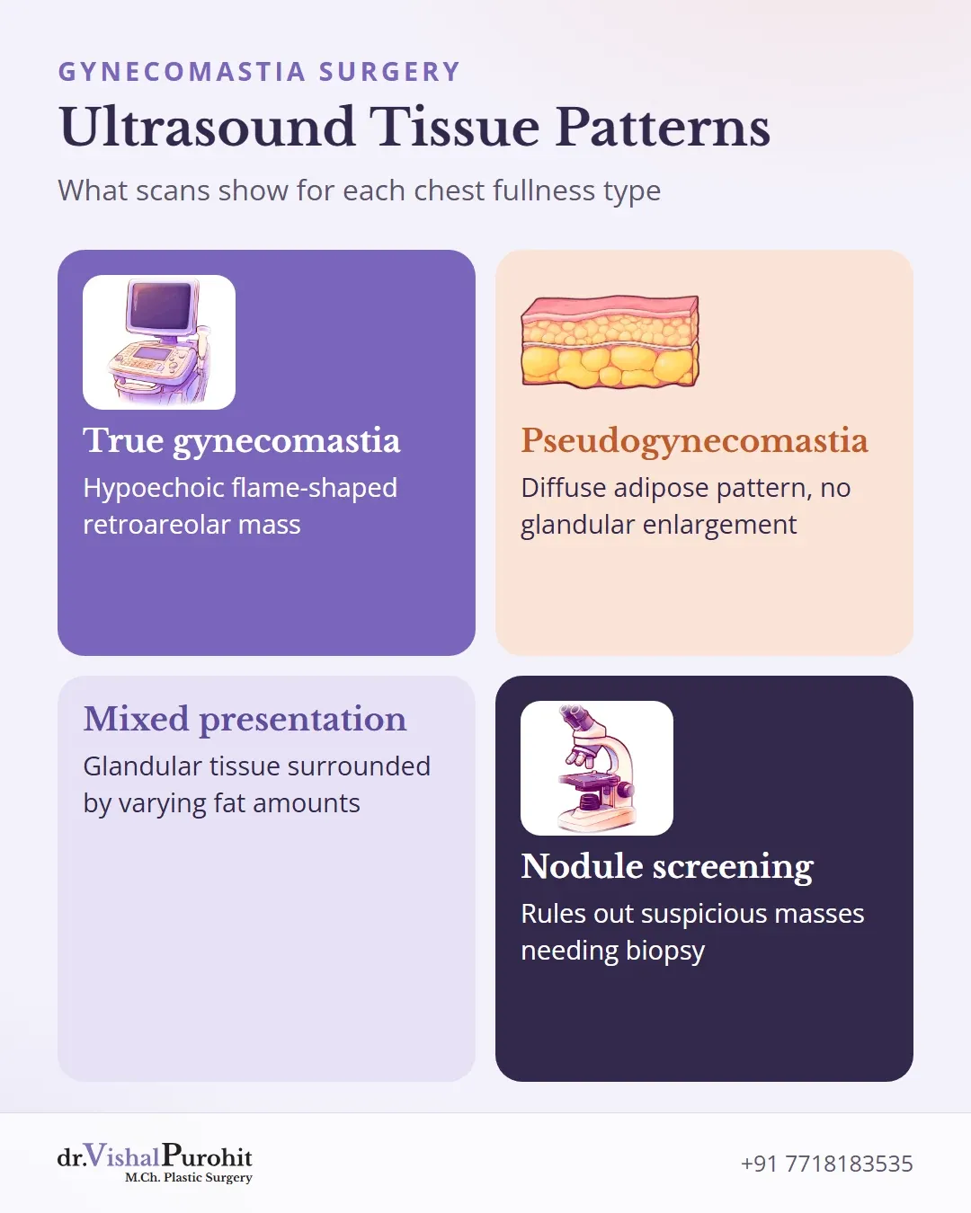
Although gynecomastia is identified clinically, ultrasound is an established initial imaging choice to evaluate male breast enlargement. It helps confirm whether breast enlargement is predominantly fatty, glandular, or a mixed combination of both tissues.
Ultrasound also differentiates tissue patterns and rules out unusual masses:
- True gynecomastia typically presents with a hypoechoic, flame-shaped retroareolar mass representing enlarged glandular tissue.
- Pseudogynecomastia displays a diffuse adipose pattern of breast fat without significant glandular enlargement.
- Mixed presentations demonstrate glandular tissue surrounded by varying amounts of fat.
In addition to identifying suspicious nodules that warrant closer review, an ultrasound objectively confirms the prevailing tissue. This verification assists the surgeon in planning targeted treatment, such as combining liposuction with direct glandular excision when true glandular tissue is present.
Why the Difference Changes Treatment
Establishing whether chest enlargement stems from adipose tissue or dense breast parenchyma determines the correct management approach.
Key points
- The type of chest tissue (fat vs. gland) dictates whether weight loss or surgery is needed.
- Weight loss reduces fat but rarely removes firm glandular tissue in true gynecomastia.
- True gynecomastia usually needs excision of the gland, while pseudogynecomastia may be treated with liposuction alone.
Weight Loss and Lifestyle Measures
For pseudogynecomastia, weight loss and structured exercise may decrease breast prominence as chest fat reduces. However, when true gynecomastia involves fibrotic or glandular parenchyma, diet and physical activity alone rarely eliminate the firm tissue. Exercise does not reverse glandular enlargement, whose causes are usually hormonal. Addressing chest fat through weight loss can reduce overall volume, but surgical excision may still be required if firm gland remains. The effect of weight change is covered in gynecomastia and weight loss.
Surgical Correction
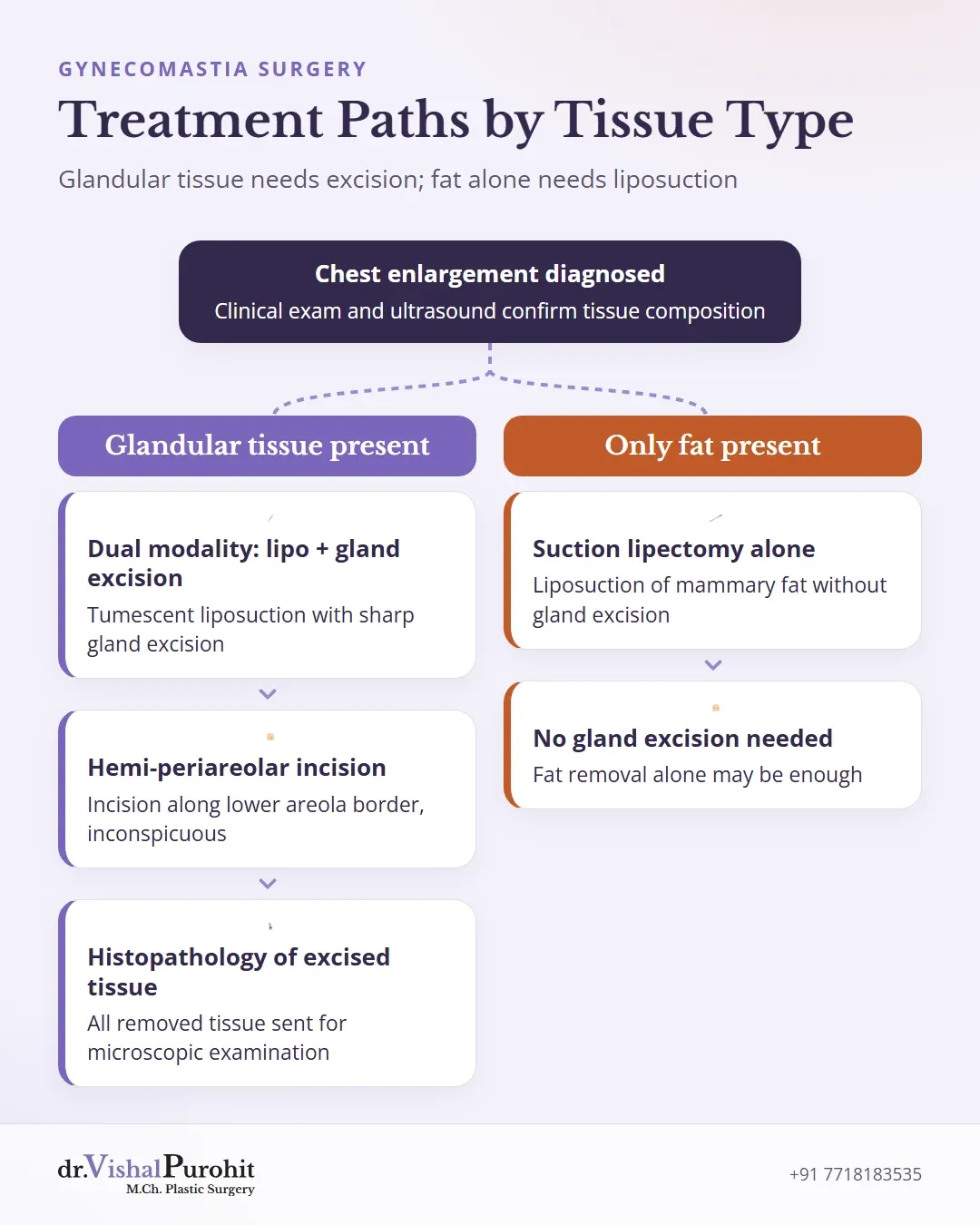
The underlying tissue composition directly guides surgical planning:
- Pseudogynecomastia: In cases characterized purely by fatty excess without glandular proliferation, suction lipectomy alone of the mammary fat is typically utilized.
- True Gynecomastia: When dense glandular hypertrophy is present, liposuction alone is often insufficient. The fibrotic glandular component is only partially cleared with suction and requires direct surgical excision.
In clinical practice in Jaipur, Dr. Vishal Purohit utilizes a dual-modality protocol combining tumescent liposuction with sharp gland excision. The glandular tissue is excised through an inconspicuous hemi-periareolar incision positioned along the lower half of the areola border; the options are compared in gynecomastia surgery techniques. Furthermore, all excised breast tissue is routinely sent for histopathology examination to evaluate the specimen.
Appropriate treatment selection depends on whether medical weight-loss management or dedicated surgical techniques are required to restore chest contour. Individual anatomy dictates whether targeted fat reduction or direct tissue removal is necessary.
Clinical Evaluation and Chest Contouring in Jaipur
Accurate identification of glandular tissue versus chest fat ensures that each patient receives the appropriate corrective strategy. To explore surgical options and aftercare details, review the guide on gynecomastia surgery in Jaipur. More answers on gland and fat are in the gynecomastia FAQs.
This is educational information, not medical advice.