
A swollen or tender chest in a man raises two questions at once: what is causing it, and is it something more serious than gynecomastia? Gynecomastia diagnosis is how a clinician answers both. In Dr. Vishal Purohit’s practice, evaluation begins with a clinical examination and direct palpation first; ultrasound or MRI is used when imaging is needed, and hormone blood tests are ordered only when there are other symptoms of hormonal imbalance. This guide, reviewed by Dr. Vishal Purohit, M.Ch. plastic surgeon in Jaipur, explains what each step looks for and why the work-up is tailored rather than the same for everyone.
An accurate assessment from a qualified M.Ch gynecomastia surgeon in Jaipur helps distinguish true glandular enlargement from pseudogynecomastia or underlying medical conditions before considering any procedural management.
If you are still at the stage of wondering what gynecomastia is, start with the overview of gynecomastia causes, symptoms and grades.
Why diagnosis comes first
Men come to a consultation for different reasons. Some notice breast pain, some feel embarrassed, and some are worried about breast cancer; in others, gynecomastia is found on a routine examination and causes no distress at all1.
Understanding which of these applies helps direct what happens next1.
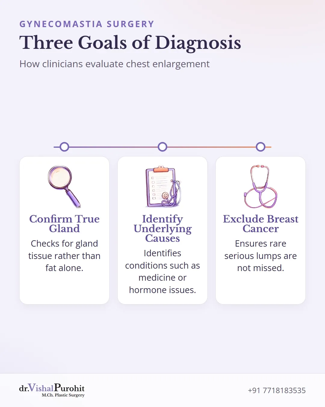
Diagnosis has three jobs:
Confirm true gynecomastia
Confirm that it is true gynecomastia, meaning enlarged gland tissue, rather than fat alone (pseudogynecomastia)2.
Look for underlying causes
Look for an underlying cause that may need treatment in its own right, such as a medicine, a hormone problem, or liver, kidney or thyroid disease1.
Exclude breast cancer
Exclude male breast cancer, which is much rarer than gynecomastia but can also present as a lump3.
Published guidance is clear that a diagnosis of “physiologic” (normal, hormone-related) gynecomastia should not be made until other causes have been excluded1.
Dr. Purohit’s note: In Dr. Vishal Purohit’s practice, evaluation begins with clinical examination and direct palpation first, followed by ultrasound or MRI when imaging is needed, while hormone blood tests are reserved only for patients with other symptoms of hormonal imbalance.
History: the questions that point to a cause
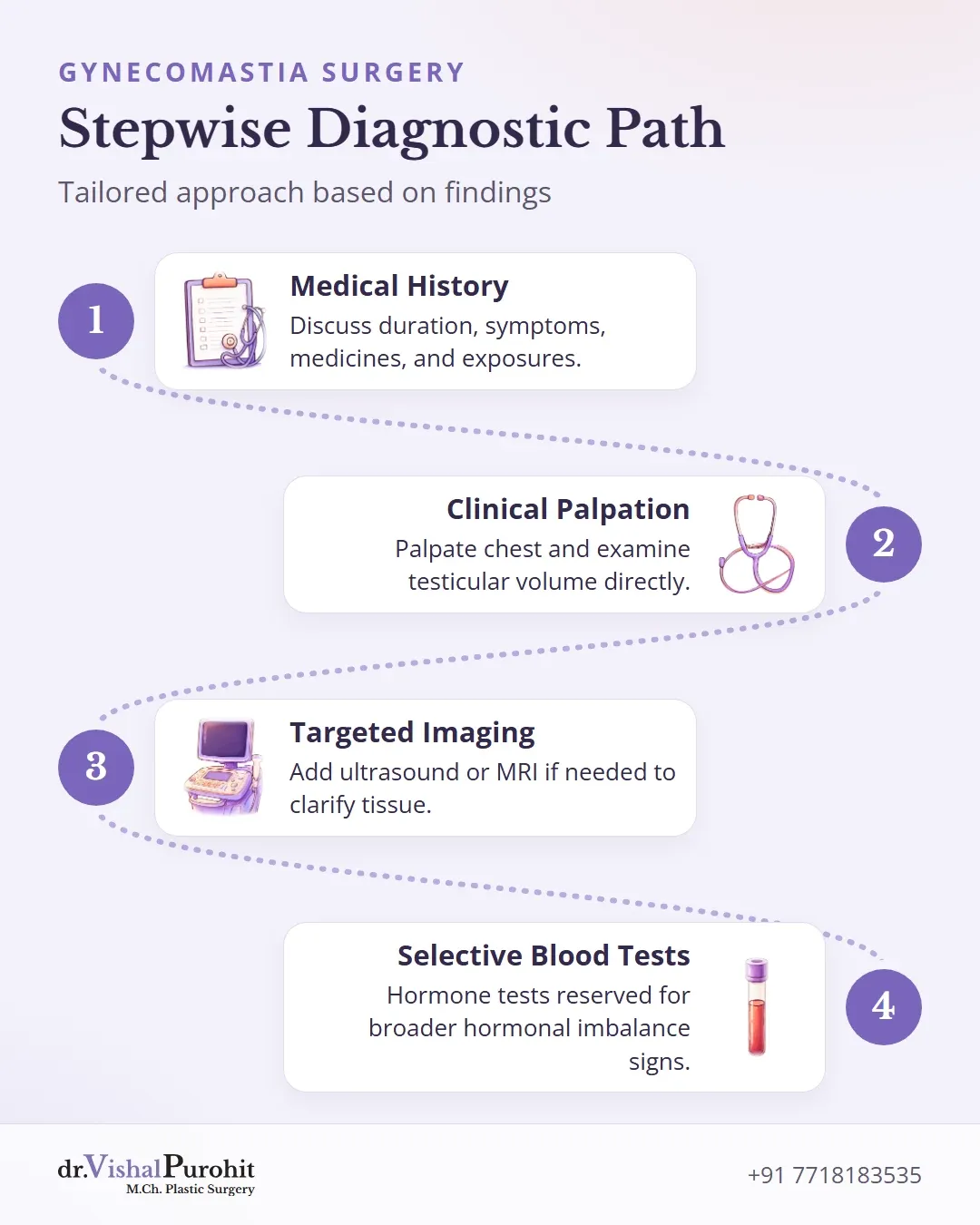
The conversation is often the most useful part of the assessment. A clinician will typically ask about145:
- When the swelling started and whether it is growing. Symptoms lasting longer than one to two years suggest a cause beyond normal hormonal change1.
- Medicines, supplements and recreational drugs. Physicians are advised to review all of these1. Medication or substance use accounts for an estimated 10 to 25 percent of cases1.
- Liver or kidney disease, sexual function and work or living exposures, all of which are part of a thorough history4.
- Family history, which may also be asked about5.
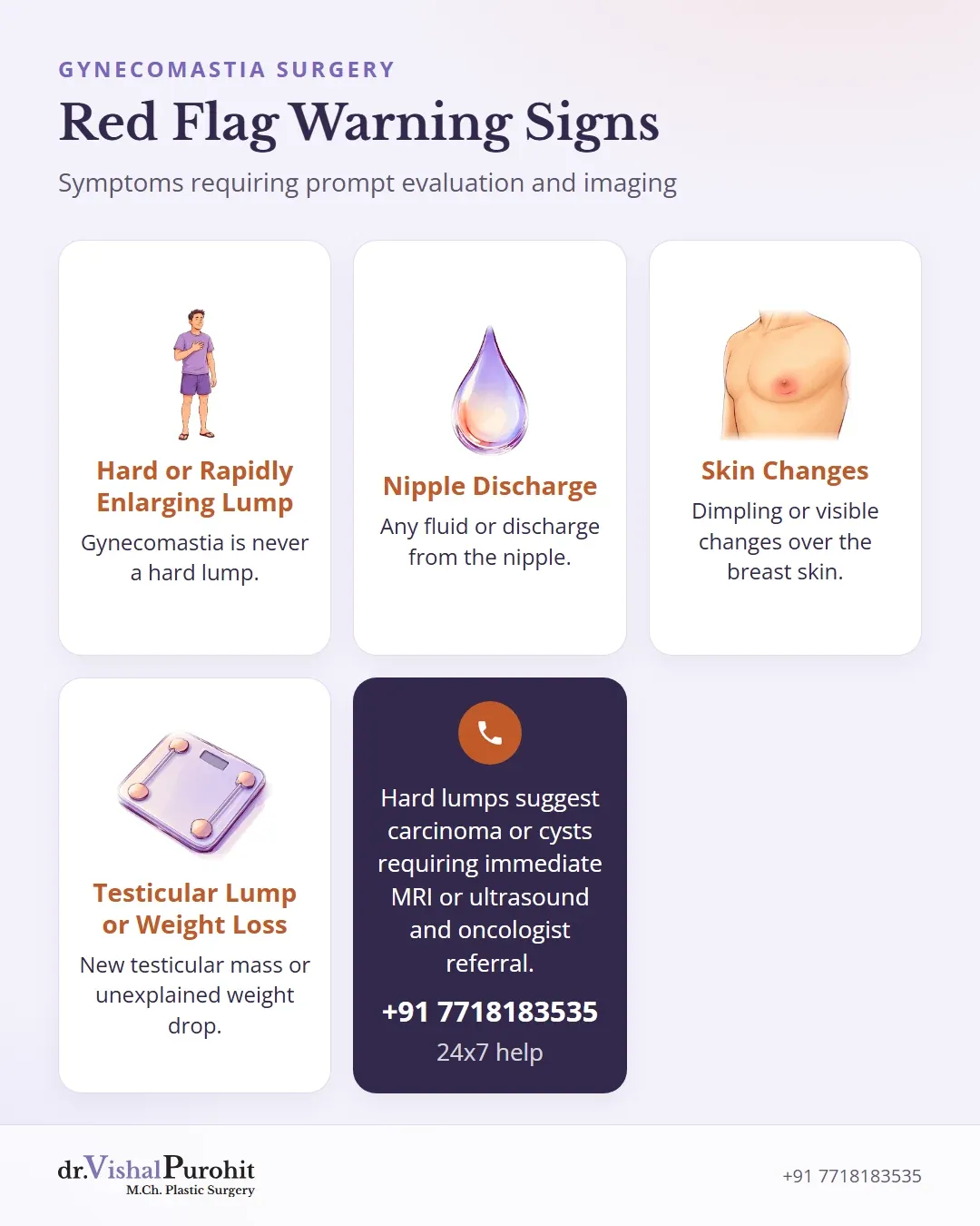
Certain answers raise concern and change the plan: nipple discharge, skin changes over the breast, a rapidly enlarging firm lump, a lump in a testicle, or general symptoms such as unexplained weight loss1. Knowing when a male breast lump should be checked helps determine if immediate medical evaluation is required.
Physical examination: gland, fat or something else

The chest
On examination, gynecomastia typically feels like a button-sized growth under the nipple that may move easily and be tender to touch; it can affect one or both sides3. In Dr. Vishal Purohit’s practice, clinical examination and direct palpation come first.
Key points
- True gynecomastia typically presents as a tender, movable glandular mass under the nipple rather than general fatty enlargement or a hard lump.
- Clinical examination assesses chest tissue, rule out cancer, and classifies findings into grades.
- Evaluation often extends beyond the chest to include testicular exams and pubertal development checks.
Gynecomastia is never a hard lump; a hard lump suggests another condition such as carcinoma or a tense cyst, which requires immediate MRI or ultrasound and referral to an oncologist. One diagnostic framework defines gynecomastia as a palpable breast mass more than 2.0 cm in diameter, and notes size, whether one or both sides are involved, discharge and tenderness4.
Telling true gynecomastia apart from pseudogynecomastia on palpation is described as a key clinical skill2. Fat-related enlargement looks like a general increase in breast size rather than a distinct lump3. The examination also looks for features of breast cancer13, noting which grades of gynecomastia match the clinical findings.
Beyond the chest
The examination may extend to the genitals and abdomen5. Dr. Vishal Purohit orders hormone blood tests only when there are other symptoms of hormonal imbalance, and refers a patient to an endocrinologist only when gynecomastia develops suddenly, well after puberty is complete, or when other symptoms of hormonal imbalance are present.
A complete assessment includes24:
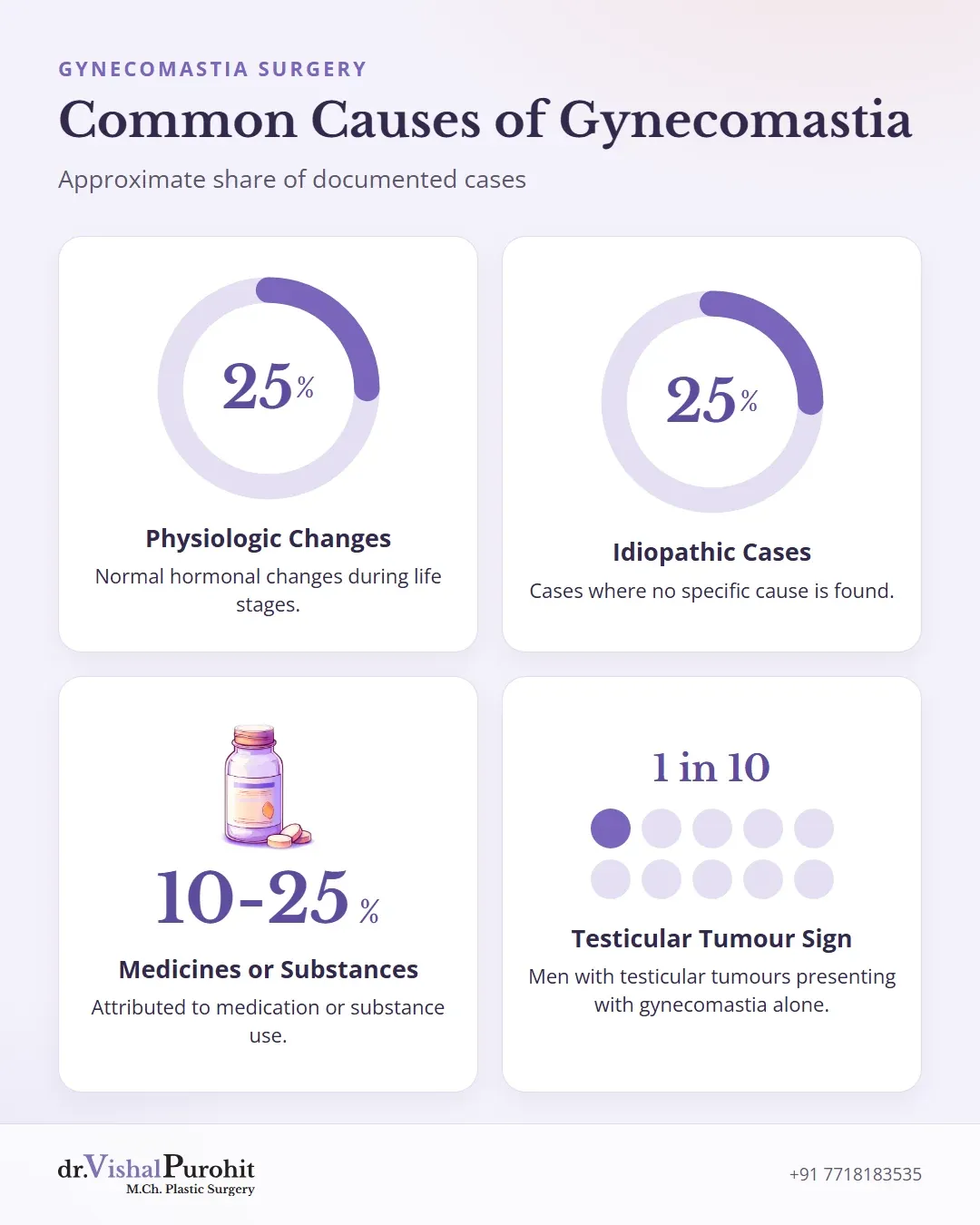
- Testicular examination, including testicular volume, to look for a mass. Testicular tumours are rare, but about 10 percent of men with testicular tumours present with gynecomastia alone1.
- Pubertal development (Tanner staging) in younger patients2.
- Secondary sexual characteristics, which are evaluated as part of a comprehensive examination4.
When hormone and other blood tests may be considered
In Dr. Vishal Purohit’s practice, clinical examination and direct palpation come first. Imaging such as ultrasound or MRI is used when needed, and hormone blood tests are ordered only when there are other symptoms of hormonal imbalance.
In the broader medical literature, blood tests are chosen from the history and examination rather than ordered as a fixed list1. Laboratory studies are selected through a stepwise approach guided by clinical findings1.
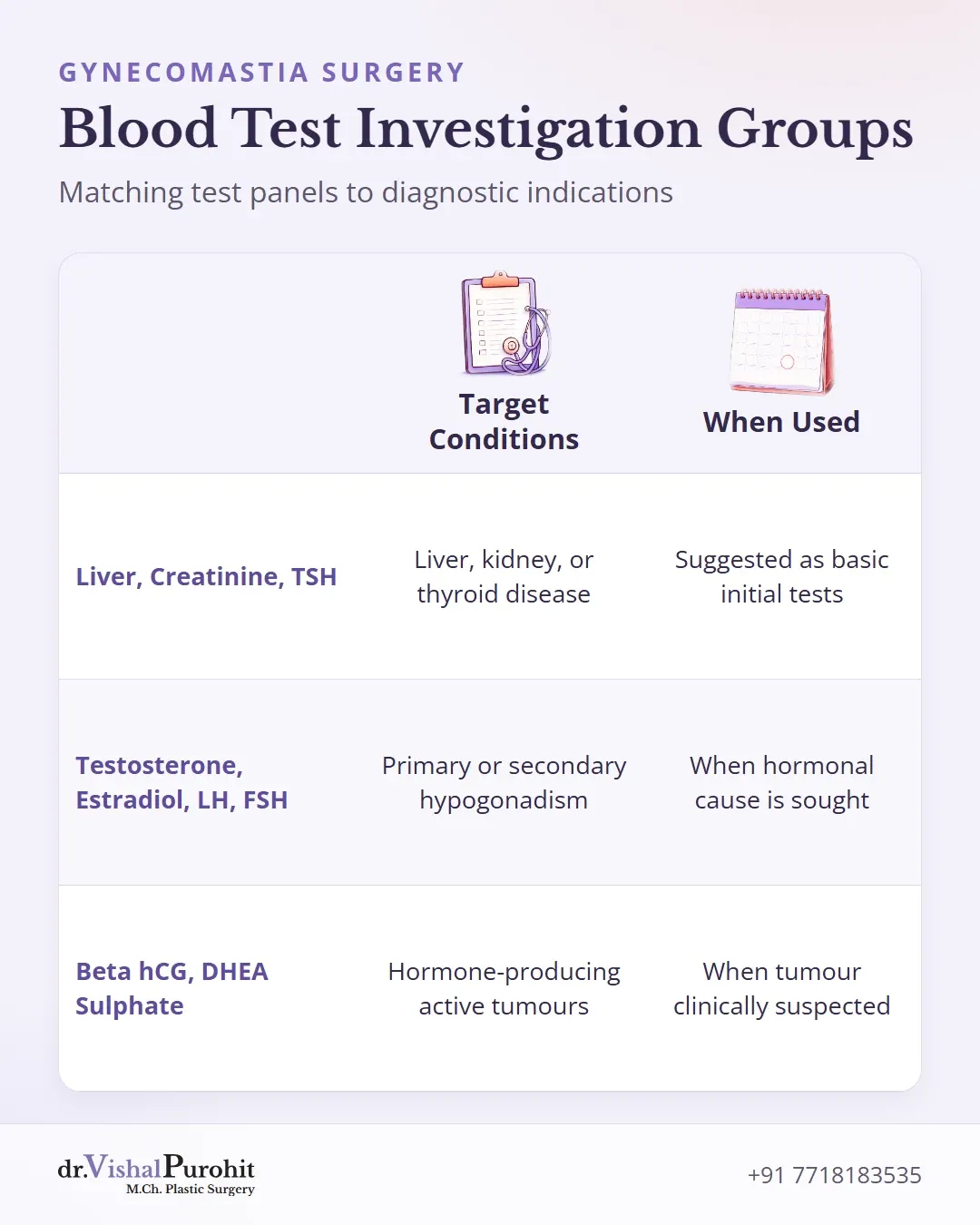
American Family Physician describes the following groups of tests1:
| Test group | What it looks for | When it is used |
|---|---|---|
| Liver enzymes (transaminases), serum creatinine, TSH | Liver disease, kidney disease, thyroid overactivity | Suggested as basic tests in the evaluation1 |
| Total and free testosterone, estradiol, LH, FSH | Hormonal imbalance from primary or secondary hypogonadism | When a hormonal cause is being looked for1 |
| Serum beta hCG, DHEA sulphate (or urinary 17-ketosteroids) | Testicular, adrenal or other hormone-producing tumours | When a tumour is clinically suspected1 |
High prolactin is not common in men with gynecomastia1. In selected cases, other tests such as a karyotype (chromosome test) for suspected Klinefelter syndrome may be considered2.
Cedars-Sinai also lists urine tests among possible investigations6.
To put these tests in context, American Family Physician summarises the approximate share of gynecomastia due to each cause1:
| Cause | Approximate share of cases |
|---|---|
| Physiologic (normal hormonal change) | 25% |
| Idiopathic (no cause found) | 25% |
| Medication or substance use | 10 to 25% |
| Cirrhosis | 8% |
| Primary hypogonadism | 8% |
| Tumours | 3% |
| Secondary hypogonadism | 2% |
| Hyperthyroidism | 2% |
| Chronic kidney insufficiency | 1% |
These blood tests look for a cause of gynecomastia. They are different from routine fitness assessments evaluated during gynecomastia surgery eligibility and preparation.
When ultrasound or mammography may be appropriate
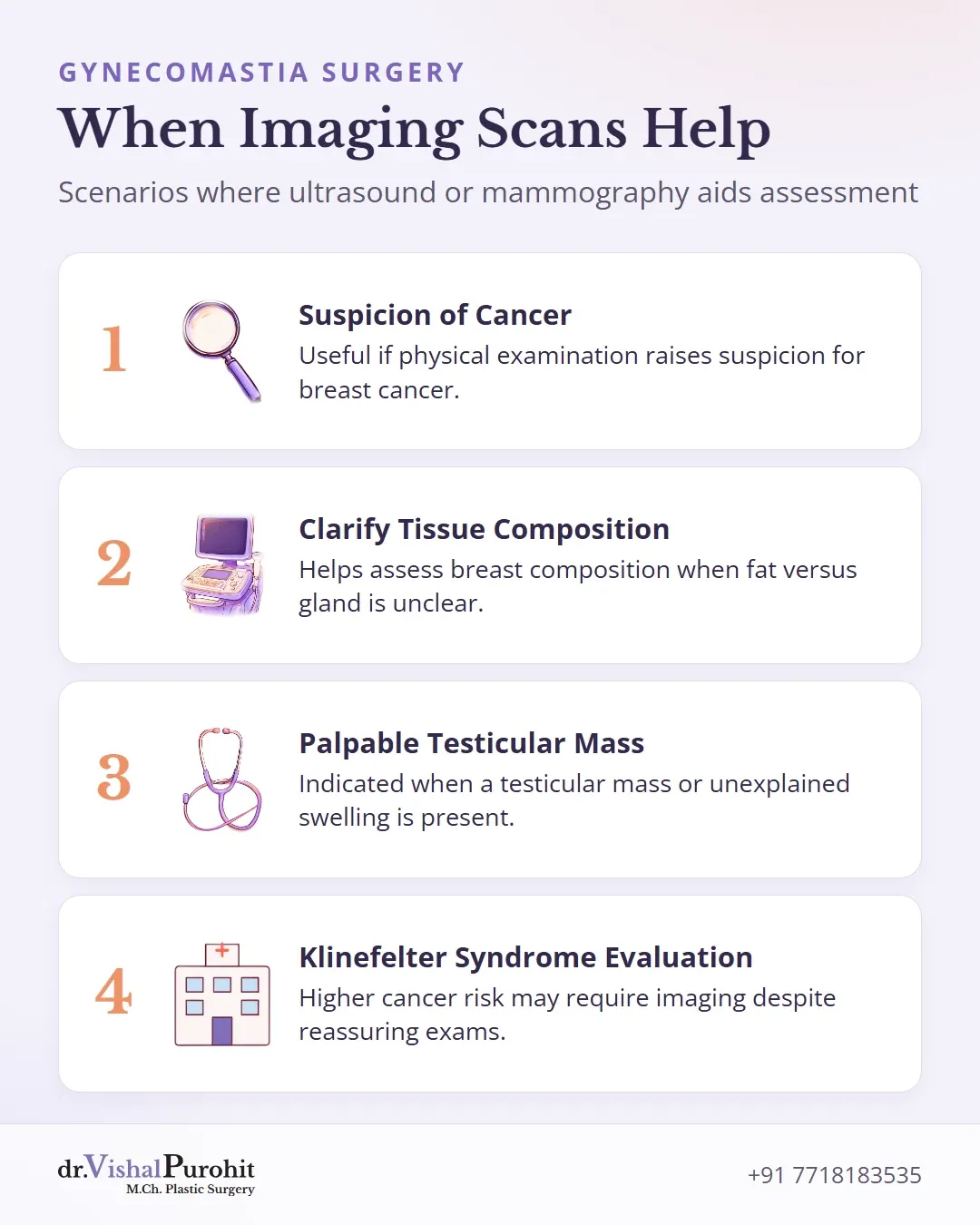
Breast imaging should be guided by the examination1. Recommendations for imaging rest mainly on case reports and expert opinion, so judgement plays a large part1.
In Dr. Vishal Purohit’s practice, clinical examination and direct palpation come first; ultrasound or MRI is used when imaging is needed.
- Breast ultrasound and mammography: Useful if the physical examination raises suspicion for breast cancer, just as in women1. Cedars-Sinai lists a low-dose X-ray of the breast among possible tests6. Dr. Purohit notes that imaging also helps distinguish gynecomastia from conditions like carcinoma or a tense cyst.
- Ultrasound to clarify tissue type: When it is unclear whether the swelling is fat or gland, ultrasound can help assess breast composition2. One group of plastic surgeons describes it as a documented modality of choice for male breast enlargement7. They request bilateral breast ultrasound before surgery to pick up suspicious nodules and confirm whether fat or gland predominates7.
- Testicular ultrasound: Some studies suggest routine testicular ultrasound to find tumours that cannot be felt1. A more widely advocated, conservative approach limits it to men with a palpable testicular mass, gynecomastia larger than 5 cm, or unexplained gynecomastia1.
- Klinefelter syndrome: Men with this condition have a breast cancer risk 16 to 30 times higher than other men and may need imaging even when the examination is reassuring1.
Imaging is generally not needed for simple obesity-related pseudogynecomastia2.
When tissue sampling or referral is needed
Most male breast lumps are benign, but breast cancer in men usually presents as a mass that cannot be told apart from gynecomastia by touch alone8. That is why sampling exists as an option.
- Fine-needle aspiration (FNA) for cytology should be pursued only if malignancy is suspected1. Where it is used, FNA is described as a sensitive, specific, simple and cost-effective way to separate benign from malignant lesions, and its routine use has reduced unnecessary surgical excisions8.
- Core needle biopsy is another option when malignant change is suspected, giving tissue for histopathology4.
- A lump that is abnormally large, tender or one-sided may prompt a biopsy or blood test to rule out breast cancer5.
Referral matters too. A consulting surgeon often benefits from enlisting an endocrinologist before proceeding with surgery9, and delaying referral to endocrinology or surgery when indicated is listed as a common mistake2.
Gynecomastia may be the only sign of primary hypogonadism; about one-half of men with Klinefelter syndrome have gynecomastia1.
Dr. Vishal Purohit sends all breast tissue removed during gynecomastia surgery for histopathology, so every specimen is examined under a microscope after the operation.
Why not every person needs every test
A universal test panel sounds thorough, but it is not how gynecomastia is assessed. In Dr. Vishal Purohit’s practice, assessment begins with clinical examination and direct palpation first.
Key points
- Gynecomastia assessment starts with physical examination rather than a standard panel of tests.
- Imaging and hormone tests are reserved for specific findings, age groups, or warning signs.
- Two people with similar appearances may require completely different evaluation steps.
Ultrasound or MRI is added only when imaging is needed, and hormone blood tests are reserved for patients who have other symptoms of hormonal imbalance. The guidance points the other way:
- The history and examination direct the work-up1. Tests are added in steps, in response to findings1.
- Over-investigating is a recognised error. Over-investigating obesity-related pseudogynecomastia is listed as a common mistake, while under-investigating true gynecomastia with other signs of hormone problems is the opposite error2. Accurate differentiation avoids unnecessary tests for benign conditions while catching the causes that matter2.
- Age changes the picture. One-half of adolescent males experience gynecomastia, typically starting at 13 to 14 years1, and in most infants and adolescents a period of observation is appropriate9. Adolescent physiologic gynecomastia should resolve within six months to two years; further evaluation is indicated if it persists beyond two years or past 17 years of age1. Outside the usual pubertal and older-age periods, clinicians keep a higher suspicion for medicines and medical causes9.
- Warning features change the plan. A rapidly enlarging firm lump, nipple discharge, skin changes, a testicular lump or weight loss move a person towards tests, imaging or sampling1. A hard breast lump is never gynecomastia; it suggests another condition such as carcinoma or a tense cyst and requires immediate MRI or ultrasound followed by referral to an oncologist.
In short, two men with similar-looking chests may have very different work-ups, and both can be correct.
How diagnosis connects to treatment planning
Before gynecomastia surgery, the surgeon evaluates several key areas:
- Discussing treatment options, goals, and medical history10
- Examining and measuring the breast tissue10
- Screening for male breast cancer and taking photographs10
Tests may be recommended both to check general health and to determine the underlying cause10. Once any correctable cause has been evaluated, the grade and tissue type guide the chosen surgical technique.
Dr. Vishal Purohit combines tumescent liposuction with gland excision through an inconspicuous hemi-periareolar incision along the lower half of the areola border. Patients can review specific details regarding recovery and costs for gynecomastia surgery in Jaipur.
In his own practice, Dr. Vishal Purohit relies on clinical examination and direct palpation first. He orders ultrasound or MRI when imaging is needed, addressing specific concerns covered in diagnosis and medical tests FAQs.
He requests hormone blood tests only when other symptoms of hormonal imbalance are present. A patient is referred to an endocrinologist only if gynecomastia develops suddenly, well after puberty is complete, or if other signs of hormonal imbalance appear.
Getting assessed in Jaipur
A clear diagnosis protects you twice: it finds a cause that may need treatment of its own, and it makes sure a lump that needs closer attention is not missed.
For most men, that means a thorough history and examination, with tests added only for a reason.
Men in Jaipur who want their chest assessed can book a consultation with Dr. Vishal Purohit, M.Ch. plastic surgeon, registered with the Rajasthan Medical Council (RMC Reg. 28041/14185), in Jaipur.
This is educational information, not medical advice.