
Botox injections for teeth grinding relax overworked masseter muscles to reduce clenching forces. Because mechanical chewing stress directly stimulates mandibular remodeling, researchers have used computed tomography scans to evaluate potential changes in bone density, volume, and cortical thickness.
While evaluating jaw pain, tooth wear and headaches: pre-Botox assessment helps identify suitable candidates, current imaging research remains limited by small cohorts and very low overall quality. Evidence does not show that observed bone variations lead to fractures, joint breakdown, or long-term clinical harm.
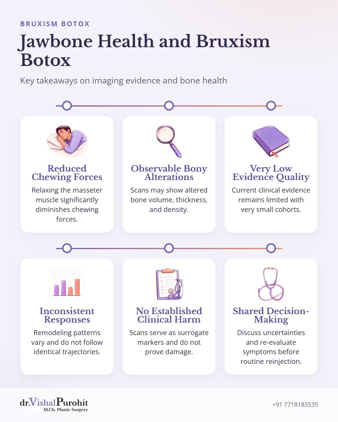
How Muscle Loading Affects Mandibular Bone
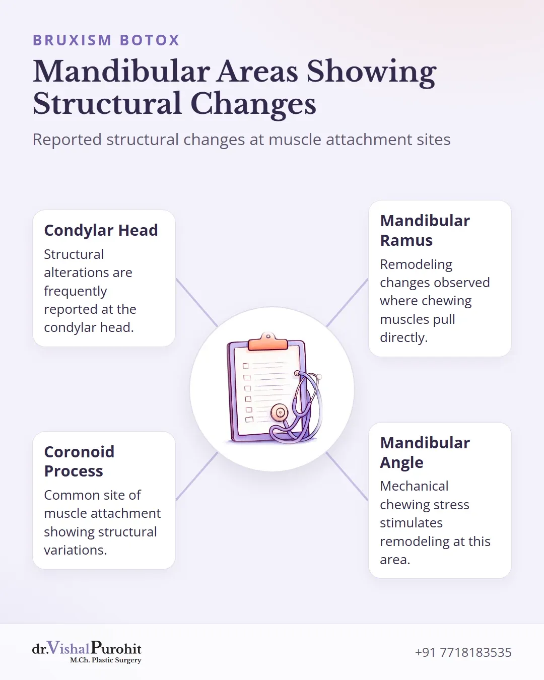
Mandibular bone maintains its density and structural shape through regular mechanical stress. Chewing muscles pull directly on bone attachments, stimulating continuous remodeling across areas like the mandibular angle, ramus, and condylar head12.
Key points
- Chewing muscles exert mechanical stress that maintains jawbone density and structural shape.
- Repeated botulinum toxin injections reduce muscle forces and may lead to localized bone resorption.
- Current studies show variable bony changes but remain limited by small sample sizes and low evidence quality.
When botulinum toxin type A relaxes the masseter muscle over repeated sessions or extended durations, it significantly diminishes these contractile forces34. This sustained drop in functional loading reduces regular mechanical stimulation, leading researchers to examine potential localized bone resorption12.
A systematic review evaluating computed tomography and cone beam computed tomography findings showed that botulinum toxin injections into masticatory muscles led to observable bony alterations in several studies1. The reported findings included:
These structural alterations were reported most frequently at the condylar head, coronoid process, and ramus1.
However, available studies remain limited by small sample sizes and very low overall evidence quality1. Interestingly, another trial in bruxism patients noted enhancements in cortical thickness at six months compared to untreated controls, demonstrating that exact bone outcomes require further long-term investigation5.
Evidence from Small Human Imaging Studies
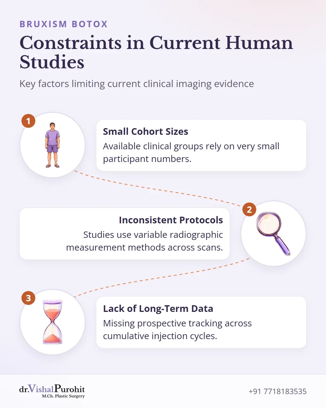
Human clinical imaging series using computed tomography (CT) and cone beam computed tomography have evaluated changes in the mandibular bone following botulinum toxin type A injections into the masticatory muscles1. These investigations assess whether muscle chemodenervation alters bone density or cortical structure at muscle attachment areas16.
Systematic reviews that examine these human CT series characterize the current clinical evidence as very low quality1. Several key constraints limit these publications:
- Very small cohort sizes across the available clinical groups1.
- Variable radiographic measurement methods and inconsistent imaging protocols1.
- A lack of robust long-term prospective data tracking cumulative injection cycles in patients16.
While experimental paralysis serves as a recognised biological model for mechanical unloading and bone loss, current clinical imaging data do not demonstrate clinically relevant bone loss in treated patients6.
Dr. Purohit’s note: Dr. Purohit reviews these imaging findings with patients to contextualise the current limits of clinical evidence during treatment planning.
Varying Findings in Recent Radiographic Research
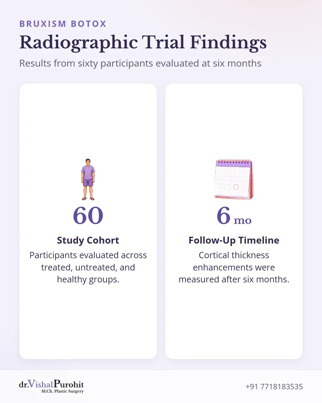
Human studies evaluating mandibular bone after botulinum toxin type A injections into the masticatory muscles using computed tomography (CT) and cone beam CT (CBCT) have examined how bone responds over time1. Recent comparative research demonstrates that these skeletal responses are not uniform across all patient groups15.
In a radiographic study assessing mandibular bone morphology in sixty participants across treated, untreated, and healthy groups, patients with bruxism who received botulinum toxin type A demonstrated:
- Substantial decreases in G2 and G3 morphological categories after six months5.
- Enhancements in cortical thickness relative to non-injected individuals and healthy controls5.
- No significant alterations in antegonial notch depth values5.
These comparative findings highlight that remodeling patterns can be subtle and divergent15. Rather than following an identical trajectory, structural adaptations to reduced muscle mechanical loading vary, demonstrating that bony responses across patient cohorts remain inconsistent15.
Why Imaging Findings Do Not Equal Clinical Harm
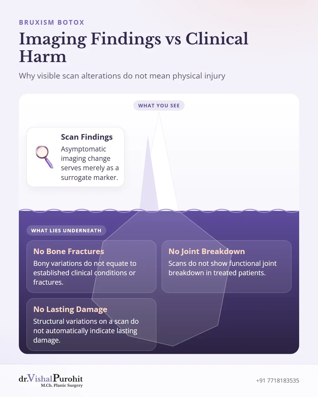
Radiological scans after botulinum toxin type A injections into masticatory muscles may reveal structural bone remodeling, such as decreased bone volume, cortical thickness, or cortical and trabecular density1. However, experimental and clinical studies indicate that evidence translating these observed structural changes into clinically meaningful skeletal harm remains limited and inconsistent6.
Key points
- Imaging may show structural bone remodeling after botulinum toxin injections, but evidence linking this to meaningful clinical harm remains limited.
- Asymptomatic radiological changes are surrogate markers that do not equate to fractures, functional breakdown, or established injury.
- Current research relies on small, low-quality studies, emphasizing the need to avoid indiscriminate multi-muscle treatment or repeated dose escalation.
Detecting an asymptomatic imaging change serves merely as a surrogate marker rather than evidence of illness. The literature confirms several key distinctions regarding these findings:
- Bony variations detected on imaging do not equate to established clinical conditions, fractures, or functional joint breakdown16.
- Demonstrating structural variations on a scan does not automatically indicate lasting damage, nor does it confirm long-term clinical safety16.
- The overall clinical significance of observed skeletal changes remains uncertain6.
Current evidence on mandibular bone quality comes from a limited number of studies with small sample sizes and very low overall quality1. While these imaging changes advise against indiscriminate multi-muscle treatment or repeated dose escalation without response6, an isolated radiological finding alone does not establish clinical injury16.
Discussing Uncertainty Before Repeat Injections
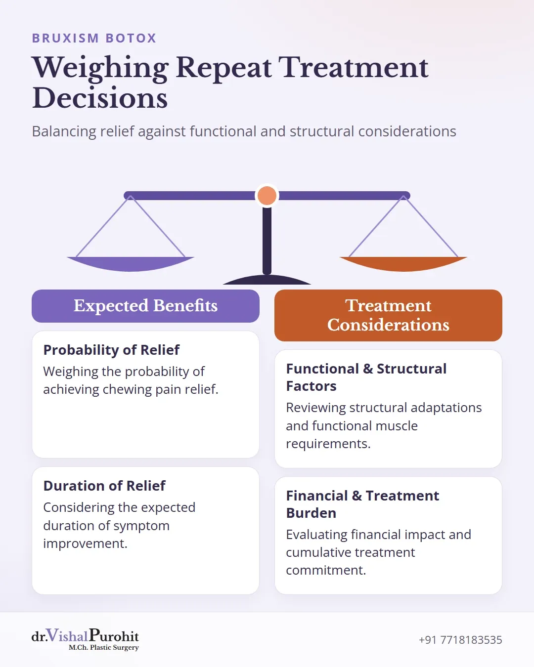
Shared decision-making is an essential part of the consultation process when evaluating ongoing care7. Clinicians and patients should discuss scientific uncertainties openly before committing to repeat injections, avoiding arbitrary maintenance schedules or unnecessary screening scans.
At the level of the individual consultation, shared decision-making should weigh the probability and expected duration of pain relief explicitly against functional, structural, financial, and treatment-burden considerations6. BoNT-A may remain a defensible option for a narrowly selected group of patients who have not responded to better-supported alternatives, whereas its routine use outside this context cannot currently be supported6.
Choosing watchful waiting or dental splints instead of routine reinjection remains a valid clinical approach. For Dr. Vishal Purohit, M.Ch. plastic surgeon, his own practice regarding repeat injections, review timing, and treatment schedules is not confirmed in advance and is decided at the consultation. Every patient is encouraged to consider alternative conservative measures alongside dental care rather than committing to fixed reinjection intervals without clinical re-evaluation.
Consultations for Bruxism Care in Jaipur
Understanding scientific evidence helps patients make informed choices regarding clenching and grinding care. Dr. Vishal Purohit, M.Ch. plastic surgeon, evaluates structural considerations alongside conservative dental management during individual consultations. Patients considering treatment can learn more about Bruxism Botox in Jaipur or read answers to common questions in the Bruxism Botox FAQs. Contact the clinic on +91 7718183535 to discuss treatment planning.
This is educational information, not medical advice.