
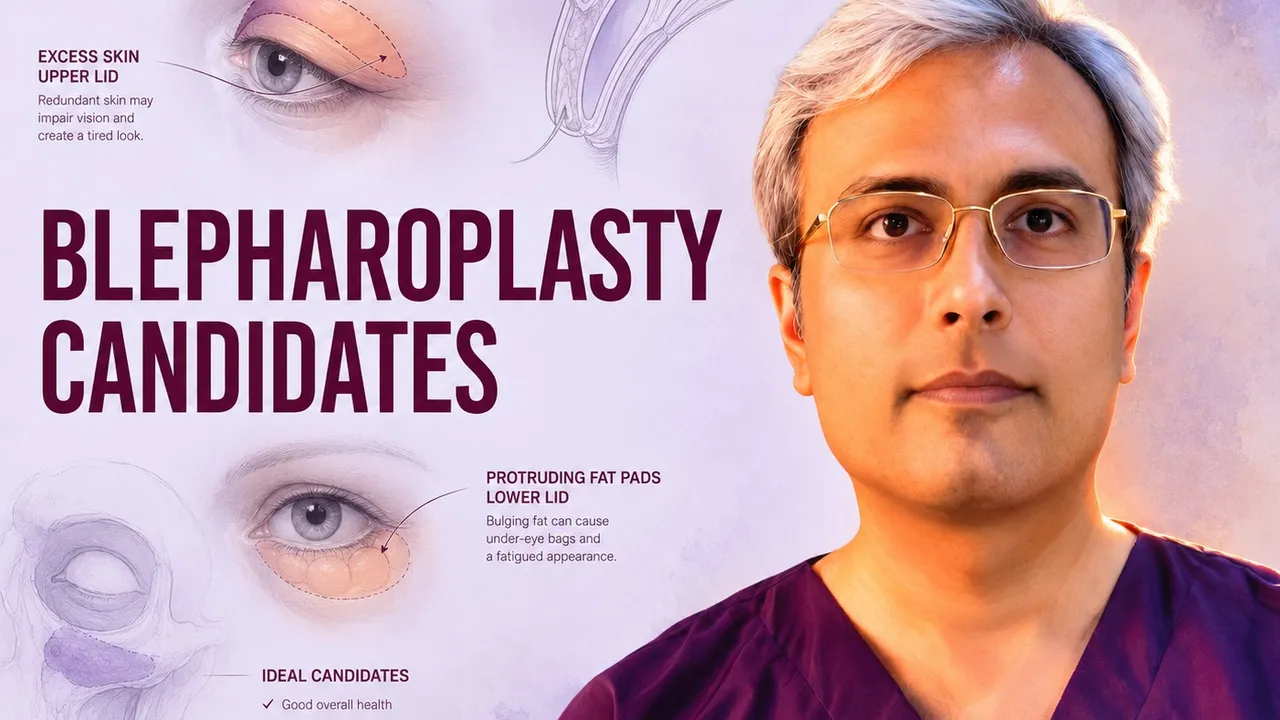
Most people who walk into a consultation for eyelid surgery have already decided what bothers them. The eyes look tired in photographs. Eyeshadow disappears into a fold. Reading gets harder by evening, and the forehead aches from a lifetime of unconsciously lifting the brows.
What they usually haven’t worked out is whether blepharoplasty is the operation that fixes it — because several very different problems produce that same tired appearance, and only some of them are treated by removing skin.
This guide covers what actually determines candidacy: the anatomy we measure, the tests that screen for trouble, who benefits most, and — just as importantly — who should wait or choose a different procedure.
At Kalpana Aesthetics in Jaipur, every plan starts from your own anatomy rather than a standard technique applied to every face. For the full picture of the procedure itself, see our complete guide to blepharoplasty.
First: Dermatochalasis or Ptosis? They Are Not the Same Operation
This distinction decides the entire surgical plan, and it is the single most common misunderstanding patients arrive with.
Dermatochalasis is redundant, lax upper eyelid skin — sometimes with prolapsed orbital fat — that drapes over the lid crease and, when severe, over the lashes. The lid margin itself sits where it should. This is what blepharoplasty treats. The operation removes a measured strip of skin, occasionally a sliver of orbicularis muscle, and conservatively reduces or repositions fat.
Ptosis is a low upper eyelid margin. The problem lies in the levator aponeurosis, its attachment to the tarsus, or Müller’s muscle — not in the skin. Blepharoplasty does not fix it. Ptosis requires a different operation: levator advancement or resection, a Müller’s muscle–conjunctival resection, or in poor-levator-function cases a frontalis sling.
Dr. Vishal Purohit explains the difference between dermatochalasis and ptosis for targeted eyelid rejuvenation. Achieve natural, youthful eyes through precise surgical correction in Jaipur.
We separate them with measurements, not impressions:
The two conditions coexist very often, and when they do, both can usually be addressed in one sitting. What we will not do is remove skin and call it ptosis correction — the lid margin would sit exactly where it started, and you would have paid for the wrong operation.
If the heaviness is true eyelid ptosis, we will tell you so at the consultation and plan accordingly.
The Brow Test That Changes Many Plans
Here is the assessment most patients have never heard of, and the one that most often changes the recommendation.
Lie a finger along the brow and hold it at its resting position, with the forehead relaxed. If the apparent excess upper lid skin largely disappears, the problem is not primarily the eyelid — it is a descended brow that the frontalis muscle has been quietly holding up, sometimes for decades.
This matters because removing eyelid skin in compensated brow ptosis makes things worse. Once the lid feels lighter, the forehead stops working, the brow drops, and the heaviness returns — now with a shortened eyelid and, in aggressive cases, difficulty closing the eye fully.
The right answer in these patients is a brow lift, either alone or combined with a conservative blepharoplasty. Recognising it beforehand is what separates a satisfying result from a revision.
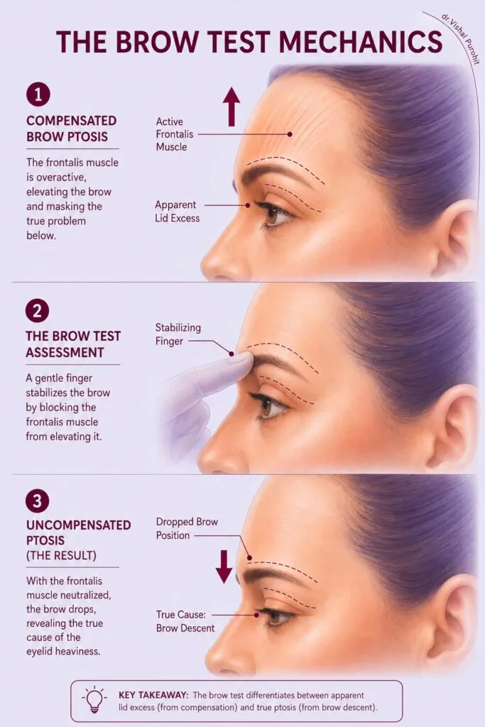
Dr. Vishal Purohit explains the brow test mechanics to differentiate between apparent lid excess and true brow ptosis. This diagnostic approach ensures precise surgical planning for optimal eyelid rejuvenation results.
Assessing Anatomical Suitability: Skin, Fat, and Muscle
Beyond the brow, the upper lid assessment looks at three layers, because each behaves differently.
Skin laxity is judged by a pinch test at the lid, taking up redundant skin with fine forceps until the lashes just begin to evert. That endpoint tells us the maximum safe excision. We also confirm at least 20 mm of skin will remain between the brow and the lid margin — go below that and lagophthalmos, incomplete closure, becomes a real risk.
Orbital fat is assessed with gentle pressure on the globe, which makes prolapsed medial and central fat pads visible. Modern practice preserves or repositions fat rather than removing it freely; a hollowed, skeletonised upper lid is one of the hardest complications to undo.
Orbicularis muscle bulk and tone are noted, along with the position of the lacrimal gland, which occasionally prolapses into the lateral upper lid and is mistaken for fat. It needs resuspension, not excision.
Understanding the layered surgical anatomy and its danger zones is what keeps dissection in the right plane.
You can read more about what happens during eyelid surgery and how different eyelid shapes affect the overall face.
A note on Asian and South Asian eyelids: crease anatomy varies considerably, and a fold that is low, absent, multiple, or asymmetric is a normal variant rather than a defect. The goal is a crease that suits your face — not a Western template imposed on it.
Lower Eyelid Candidacy: A Separate Assessment
Lower lid surgery addresses different complaints — fat pads producing under-eye bags, a visible tear trough, and skin crepiness — and it carries different risks. Candidacy turns on lid support.
Snap-back test. The lower lid is drawn down and released without blinking. A healthy lid returns to the globe immediately. A slow return signals laxity.
Distraction test. The lid is pulled forward from the globe. More than 6–8 mm of distraction indicates significant laxity.
Vector assessment. Viewed in profile, if the cheek’s most anterior point sits behind the cornea — a negative vector — the eye is relatively prominent, and routine lower blepharoplasty carries a higher risk of scleral show and lid retraction.
Lax or negative-vector lower lids are not a bar to surgery, but they change it: a canthopexy or canthoplasty is added to support the lid, and skin excision becomes far more conservative. Patients who are told about this beforehand do well. Patients who are not are the ones who develop rounded lids and chronic irritation.
Where the concern is purely a hollow tear trough with good skin quality, fat repositioning or filler may serve better than any excision at all.
Functional vs. Aesthetic Goals: Which Is Driving You?
Both are legitimate reasons to operate, and most patients have a mix. Naming the dominant one shapes the plan.
Functional candidacy is objective and documentable. Superior visual field testing quantifies the loss caused by overhanging tissue, and a meaningful proportion of patients show measurable visual field improvement after upper lid surgery. Chronic head tilt and constant brow elevation produce genuine occipital and frontal strain. A deep, moist fold that macerates and reddens is a dermatological problem, not a cosmetic one.
Aesthetic candidacy is subjective, and that is fine — wanting to look less tired is a reasonable thing to want. What we look for is a goal that is specific and proportionate. “I want my eyeshadow to show again” is a workable brief. “I want to look like a different person” is not, and neither is a complaint that is invisible to everyone except the patient. Where the concern seems out of proportion to the finding, or where the patient has had multiple procedures each of which failed to satisfy, we slow down and talk rather than operate; patient expectations predict satisfaction more reliably than any anatomical variable.
You May Be a Strong Candidate If…
-
Upper lid skin rests on or over the lashes, or a fold obscures the natural crease
-
Superior visual field is measurably reduced, or you catch yourself lifting your brows to read
-
The brow sits at a normal height and does not account for the heaviness
-
Lower lid fat pads are prominent with reasonable lid tone and skin quality
-
Ocular surface is healthy, or a dry eye is well controlled on treatment
-
General health is stable, non-smoking or willing to stop, and any blood pressure, thyroid, or diabetes is under control
-
Expectations are specific, realistic, and your own — not someone else’s suggestion
-
You can protect two weeks for visible recovery
You May Not Be a Good Candidate — At Least Not Yet
This section is as important as the one above, and an honest surgeon says it out loud.
An informative guide on blepharoplasty candidacy and screening requirements. Dr. Vishal Purohit ensures patient safety and optimal surgical results through comprehensive pre-operative evaluation.
Significant dry eye or ocular surface disease. Blepharoplasty transiently worsens tear film function, and pre-existing dry eye can become genuinely miserable afterwards. We screen with symptom history, Schirmer’s test, tear break-up time, and corneal staining. Dry eye is not an absolute bar — it means treat first, operate conservatively, and set expectations. It is never something to discover post-operatively.
Absent or weak Bell’s phenomenon. If the globe does not roll up on lid closure, the cornea has less protection during any period of incomplete closure. Skin excision must be markedly more conservative.
Active or unstable thyroid eye disease. Lid retraction and proptosis in Graves’ orbitopathy mimic the appearance blepharoplasty treats, and operating on active disease produces unpredictable results. The disease should be inactive and stable for at least six months, and any orbital decompression or strabismus surgery must precede lid surgery.
Recent refractive surgery. LASIK reduces corneal sensation and tear production for several months. We generally wait six months and confirm a stable ocular surface.
Uncontrolled systemic disease. Poorly controlled hypertension raises the risk of the one complication that genuinely threatens vision: retrobulbar haemorrhage. Uncontrolled diabetes impairs healing. Bleeding disorders and anticoagulation need planning with your physician — never independent stopping of your own medication.
Smoking. Nicotine compromises flap perfusion and healing. We ask for cessation at least four weeks before and four weeks after surgery.
Uncontrolled glaucoma, or a history of retinal detachment or eye trauma — these require ophthalmology clearance first.
Unrealistic expectations, or surgery motivated by someone else. A patient whose distress is disproportionate to the physical finding, or who is pursuing surgery to save a relationship or a job, is not helped by an operation. Recognising this is part of the assessment, not a judgement of the patient.
Pregnancy or breastfeeding, and any period of significant emotional upheaval. Elective surgery can wait.
Age, Skin Elasticity, and How Long Results Last
Age alone neither qualifies nor disqualifies anyone. Candidacy in the twenties and thirties is usually about inherited fat pads or a low crease rather than aging; the operation is conservative and largely fat-focused. From the forties onward, skin redundancy becomes the dominant issue and excision plays a larger role. Beyond sixty, brow position, lid laxity, and ocular surface health matter more than the calendar, and a combined brow-and-lid approach becomes more common.
Skin quality — elastic recoil, sun damage, thickness — predicts how well the tissue redrapes and how the scar will settle. It influences technique choice more than any single number.
On longevity, an honest answer: upper blepharoplasty results are long-lasting, and many patients never need a second procedure, though aging continues and some skin laxity may return over ten to fifteen years. Lower lid fat removal is generally permanent, since the fat pads do not regenerate. Neither operation stops the aging process — it resets the starting point.
Medical History and Surgical Safety Evaluation
We discuss the risks and safety realities of eyelid surgery openly at consultation, including the rare ones. A patient who understands what can go wrong is a safer patient.
What Recovery Actually Looks Like
Recovery is where expectations most often diverge from reality, so here is the honest timeline.
Temporary dryness, grittiness, watering, and slight blurring from ointment are normal in the early weeks. Sun protection over the scars for six months materially improves how they settle.
Tailoring Results to Your Eye Shape
There is no single ideal eyelid, and results that look obviously “done” almost always come from applying one to every face. Crease height and shape, degree of medial fold, the eye’s axial tilt, brow-to-lid distance, and the way the lid relates to the cheek and midface all vary — and all vary further across ethnicities.
Planning is conservative by default, and asymmetry is measured rather than assumed, because almost every face is naturally asymmetric and patients frequently notice a pre-existing difference for the first time after surgery. Pointing it out beforehand, in photographs, avoids a great deal of post-operative anxiety, and the surgical literature on cosmetic outcomes consistently favours restraint over aggressive excision.
Frequently Asked Questions
What criteria determine if I am a good candidate for blepharoplasty in Jaipur?
An ideal candidate is an adult experiencing hooded upper eyelid skin, obstructed peripheral vision, or persistent lower eyelid fat bags, clinically evaluated by Dr. Vishal Purohit in Jaipur. Candidates should have healthy facial tissue and realistic expectations without uncontrolled dry eye syndrome or glaucoma. As a board-certified plastic surgeon, Dr. Purohit performs calibrated pinch testing to ensure safe eyelid closure.
What is the difference between upper and lower blepharoplasty?
Upper blepharoplasty excises sagging skin and herniated medial fat through a discreet incision hidden in the supratarsal crease, while lower blepharoplasty removes or repositions under-eye fat bags via a transconjunctival or subciliary incision, performed by Dr. Vishal Purohit in Jaipur. Upper surgery lifts heavy, drooping lids; lower surgery smooths dark hollows and puffiness. Dr. Purohit customizes procedures to each patient’s periorbital anatomy.
Can blepharoplasty improve obstructed peripheral vision?
Yes, functional upper blepharoplasty elevates severe dermatochalasis (overhanging skin) that droops over the eyelashes, instantly expanding the visual field and eliminating eyebrow fatigue, confirmed by Dr. Vishal Purohit in Jaipur. Many patients notice improved reading comfort and reduced forehead strain. As an M.Ch. plastic surgeon, Dr. Purohit documents visual field defects for medical clearance.
What is the recovery time and downtime following eyelid surgery?
Most patients return to desk work, driving, and social routines within 7 to 10 days following blepharoplasty by Dr. Vishal Purohit in Jaipur. Eyelid swelling and bruising peak at 48 hours and fade rapidly with cold compresses, while micro-sutures are removed painlessly at day 5 to 7. Dr. Purohit provides tailored post-operative eye drops and ointment to ensure smooth healing.
Will blepharoplasty alter my natural eye shape or make me look startled?
No, Dr. Vishal Purohit in Jaipur employs conservative skin resection and fat-preservation techniques that rejuvenate the eyes while preserving your authentic ethnic shape and natural eyelid aperture. Avoiding excessive skin removal prevents hollowed or startled expressions. Dr. Purohit’s delicate surgical aesthetic delivers an awake, refreshed, and naturally youthful gaze.
Next Step
The only way to answer the candidacy question properly is an examination — measurements, ocular surface tests, and standardised photographs, with an honest conversation about what surgery can and cannot do for your face.
To discuss your case or book a consultation, contact Dr. Vishal Purohit in Jaipur at +91-7718183535.
Medically reviewed by Dr. Vishal Purohit, Plastic and Cosmetic Surgeon, Jaipur. Last updated 25 July 2026.
This article is for general information and does not constitute medical advice or a substitute for individual consultation. Surgical results vary between individuals; no specific outcome is implied or guaranteed. Every surgical procedure carries risk, which will be discussed with you in detail before any decision.
Expert Blepharoplasty & Eyelid Rejuvenation in Jaipur
Are heavy, drooping upper eyelids or prominent under-eye bags making you look tired, aged, or obstructing your visual field? Schedule a detailed periorbital examination with Dr. Vishal Purohit, board-certified M.Ch plastic surgeon in Jaipur.
- Comprehensive Master Guide: Explore our foundational clinical overview on What is Blepharoplasty? Eyelid Surgery Explained.
- Surgical Procedures & Consultations: Visit our specialized Blepharoplasty Surgery in Jaipur suite to review surgical approaches, recovery milestones, and book your clinical evaluation.