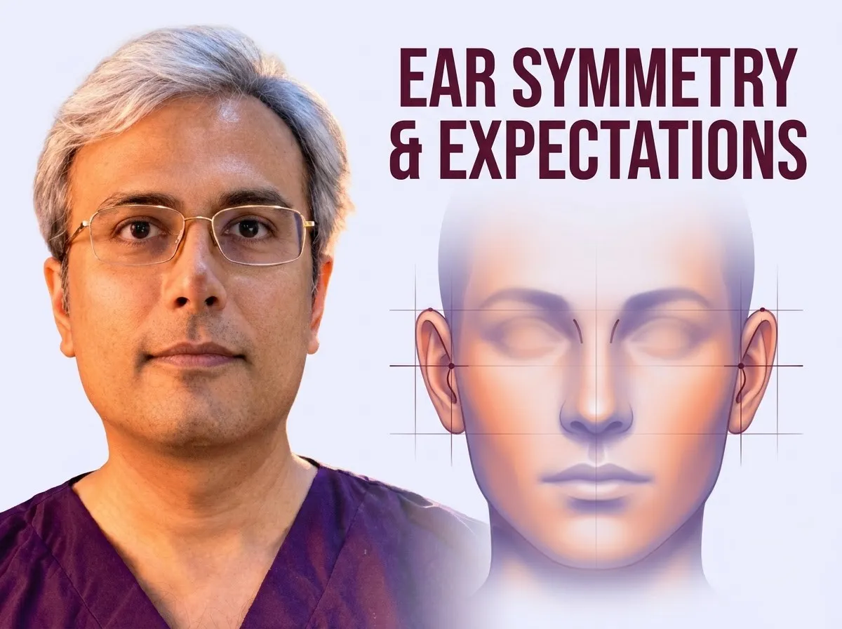
Natural ears show biological variations in height, shape, and protrusion across the general population. Because the ears sit on opposite sides of the head, absolute bilateral symmetry is neither present in nature nor expected after surgery.
Dr. Vishal Purohit assesses underlying cartilage anatomy during consultations for otoplasty in Jaipur to help patients develop balanced, realistic expectations rather than pursuing artificial millimeter-perfect matching.
Surgical correction focuses on balanced projection and gentle contours rather than identical measurements. Patients exploring surgical anatomy can read about why ears stick out, ear folds, the conchal bowl and hearing to understand how cartilage structure guides realistic surgical planning.
Two Ears Are Never Identical
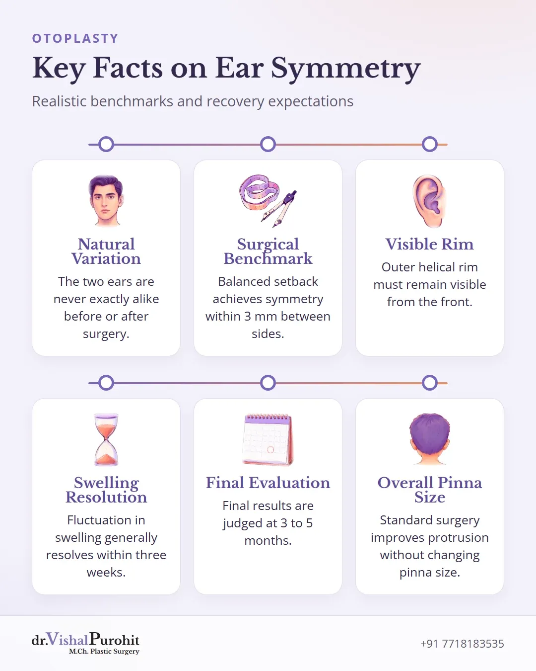
In the practice of Dr. Vishal Purohit, an M.Ch. plastic surgeon in Jaipur, patients are reminded that the natural variance between ears is entirely acceptable because nobody sees both ears fully at the same time.
Dr. Purohit’s note: Dr. Purohit reminds patients that the two ears are never exactly alike, before or after surgery, and perfect symmetry cannot be achieved.
Surgical literature confirms that mild differences between the right and left sides are normal biological variations1. The ears are situated laterally over the temporal bones, meaning their complete shape cannot be viewed simultaneously from the front2. Because an observer must look at one ear and then shift positions to inspect the contralateral side, absolute bilateral symmetry is neither naturally present nor clinically expected2.
Preoperative differences in ear dimension, shape, protrusion, rotation, and height on the head occur across the general population12. For this reason, established otoplasty surgical benchmarks typically define an aesthetically balanced outcome as achieving symmetry within 3 mm between the two sides34. The primary operative focus remains on creating a natural contour and balanced projection when viewed from the front, rather than seeking identical measurements23.
What a Natural Result Looks Like
A natural outcome aims to restore balanced contours without giving the impression that the ears have been surgically pinned back5. As Dr. Vishal Purohit notes in his practice, natural variation between the ears is acceptable because nobody views both ears fully at the same time.
Key points
- Natural results restore balanced contours without creating an artificially pinned-back look.
- The outer helical rim should remain visible from the front with smooth, rounded antihelical contours.
- Balanced suture tension across all ear segments helps avoid telephone and reverse telephone ear deformities.
Visibility of the Helical Rim
From the frontal view, the outer helical rim should remain visible67. When an ear is set back, the antihelix should have smooth, rounded contours rather than sharp or unnatural folds6. In standard aesthetics, the helix should ideally lie slightly beyond or more lateral to the antihelix67. If the setback is excessive or the antihelix over-projects, the rim can disappear behind the antihelical fold, creating a telltale sign of an operated ear78.
Avoiding the Telephone Ear Deformity
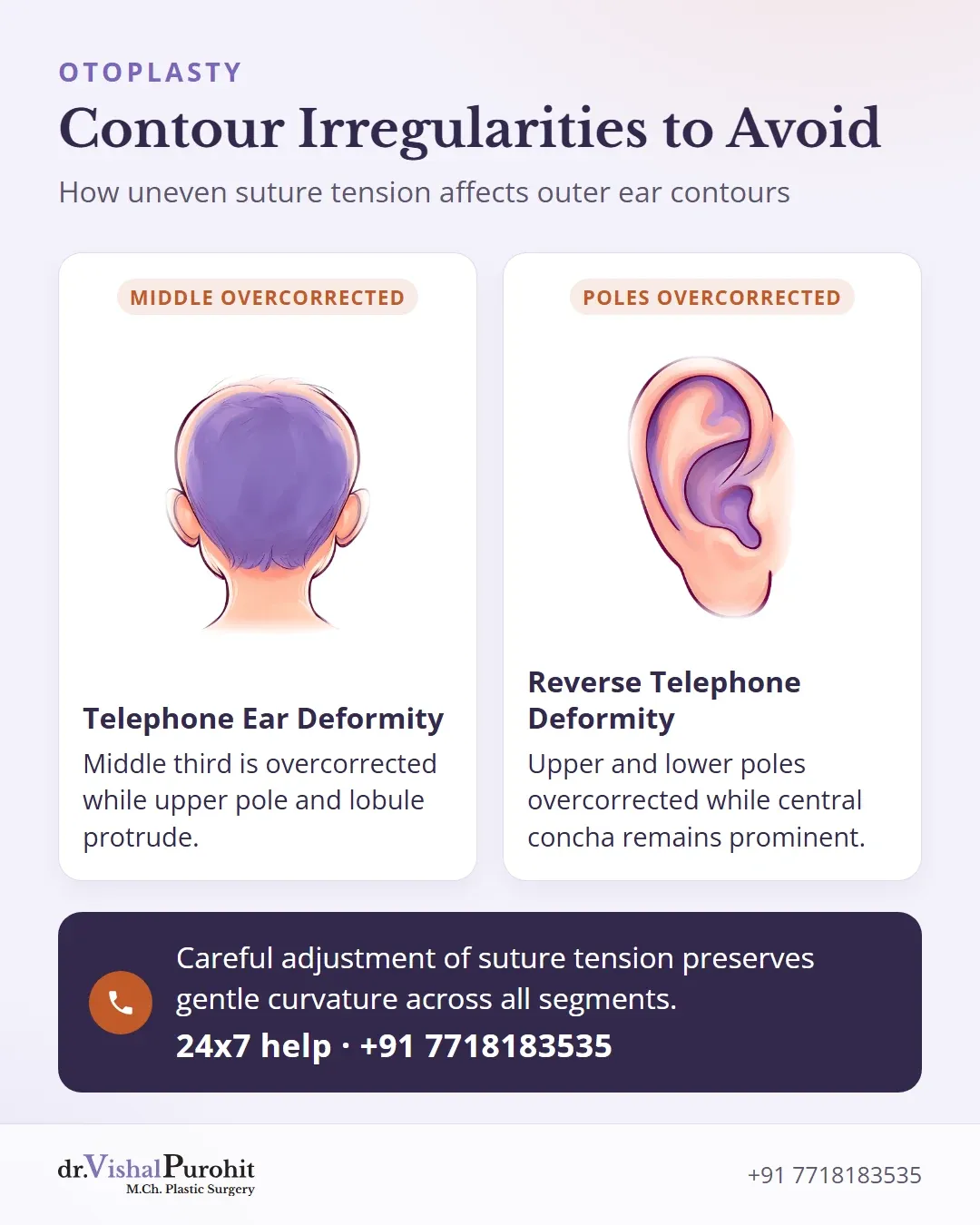
Surgical planning must also avoid contour deformities:
- Telephone ear deformity: Occurs when the middle third of the ear is overcorrected or compressed while the upper pole and lobule remain protuberant, resembling a classic telephone receiver91011. This often arises from excessive tension on central sutures, such as Mustardé or concho-mastoid sutures, or uneven tissue resection911.
- Reverse telephone ear deformity: Happens when the upper and lower poles are overcorrected while the central concha remains prominent89.
Careful adjustment of suture tension across all segments helps preserve gentle curvature and prevents a flattened appearance911.
Swelling and the Timeline
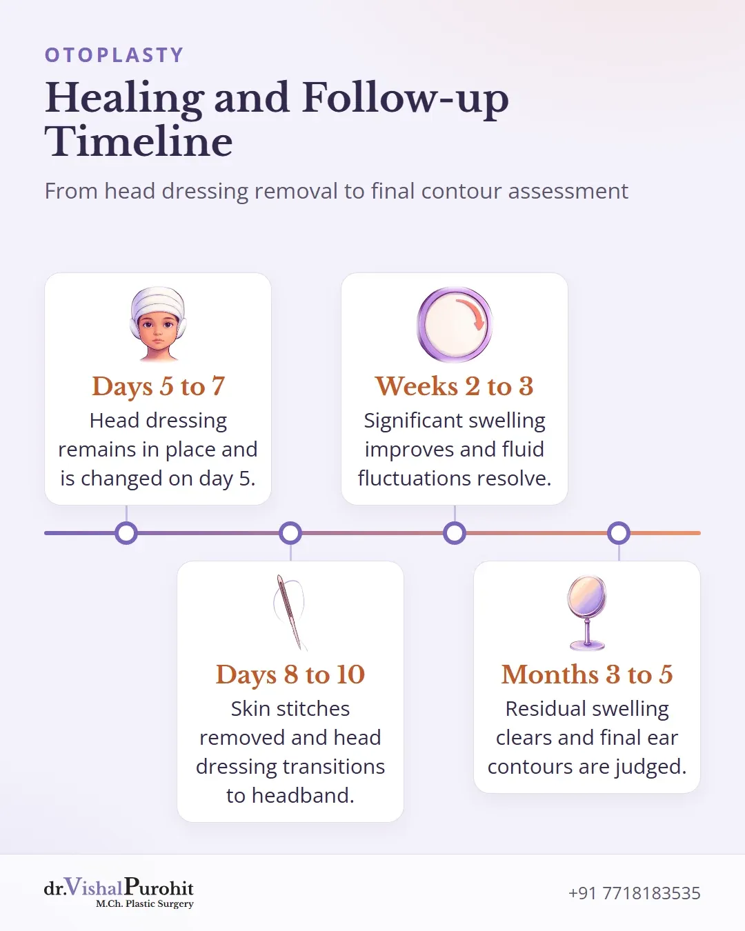
In Dr. Vishal Purohit’s practice, the final result of otoplasty is judged at 3 to 5 months.
Immediately following surgery, the initial ear shape is hidden beneath a head dressing, which remains in place for 5 to 7 days and is first changed at the day 5 review. Early tissue swelling also conceals the true contour and definition of the ears once the dressing is removed.
Literature shows that most patients experience significant improvement in swelling within 2 to 3 weeks, with mild residual oedema persisting through week 612. Fluctuation in oedema generally resolves within the first three weeks12.
To support healing after the head dressing is removed, Dr. Purohit advises patients to wear a soft headband day and night for 1 to 2 weeks, followed by night-time wear for an additional 3 to 4 weeks. Skin stitches are removed between day 8 and day 10. Formal follow-up reviews occur on day 5, day 10, at 1 month, and at 3 months to monitor healing as the residual swelling gradually clears.
What Otoplasty Does Not Change
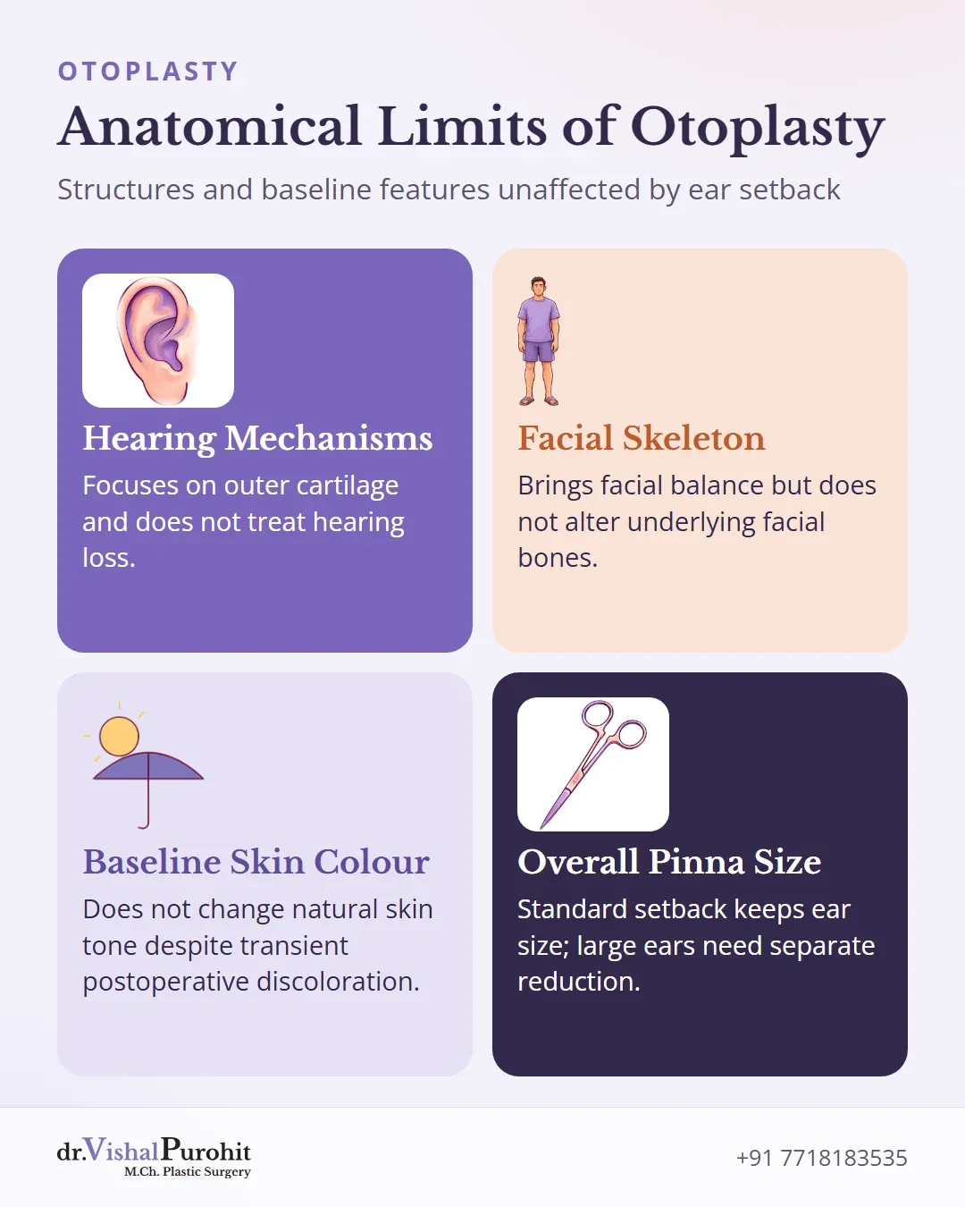
Otoplasty adjusts the structural framework of the outer ear, but it has defined anatomical limits. Understanding what the procedure cannot alter helps establish realistic expectations before surgery.
Key points
- Otoplasty reshapes the external ear cartilage but does not treat hearing loss or alter internal hearing mechanisms.
- The procedure balances ear proportion without changing underlying facial bone structure or baseline skin colour.
- Standard pinning corrects protrusion rather than overall pinna size, which requires specialised reduction techniques.
Hearing Function
Otoplasty changes the shape, position, or proportion of the external ear and is not performed to treat hearing loss13. The surgery focuses on the visible cartilage structure and does not alter internal hearing mechanisms, though operative risks can include external canal stenosis or middle ear injury3.
Facial Structure and Skin Colour
Ear surgery brings balance and proportion to the ears and face13, but it does not alter the underlying shape of the facial skeleton or facial features. It also does not alter baseline skin colour, though transient skin discoloration and swelling remain recognised surgical risks14.
Ear Size and Pinna Reduction
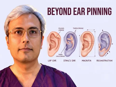
Standard otoplasty improves ear protrusion without affecting the overall size of the pinna15. Addressing overly large ears (macrotia)13 or reducing pinna dimensions requires specific reduction incisions that leave visible scars15. In Dr. Vishal Purohit’s practice, cases involving large ears are accepted for evaluation and correction.
Checking Progress at Reviews
Standard clinical photographs are useful for checking progress. Standard views for otoplasty include frontal with hair pulled back, posterior with hair pulled back, and right and left lateral views, including close-ups16.
Patients attend follow-up reviews at 1 month and 3 months to assess healing and ear shape. Any worries about balance, healing, or symmetry can be raised during these appointments. For details on earlier care, see the dressing post.
Patient Satisfaction in Studies
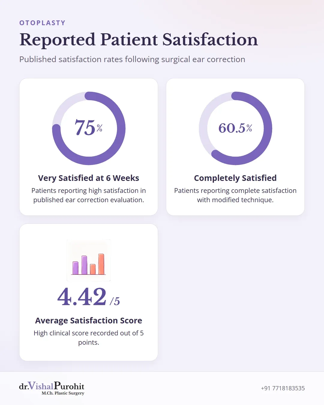
Published research shows generally high satisfaction following otoplasty1718. In one evaluation of ear correction, 75% of patients reported being very satisfied and 23% satisfied at 6 weeks18. Another study reported an average satisfaction score of 4.42 out of 5 for a modified technique19, with 60.5% completely satisfied and 29.6% mostly satisfied20. Dissatisfaction in the literature was rare and largely linked to obvious clinical recurrence17.
Consulting on Ear Correction in Jaipur
Achieving balanced ear contours requires realistic expectations, careful cartilage shaping, and patience while post-operative swelling resolves. Individuals seeking balanced correction can explore options for otoplasty in Jaipur to address uneven or prominent ears. For further clinical guidance on dressings, recovery precautions, and surgical planning, read the detailed otoplasty FAQs before scheduling a clinical evaluation.
This is educational information, not medical advice.