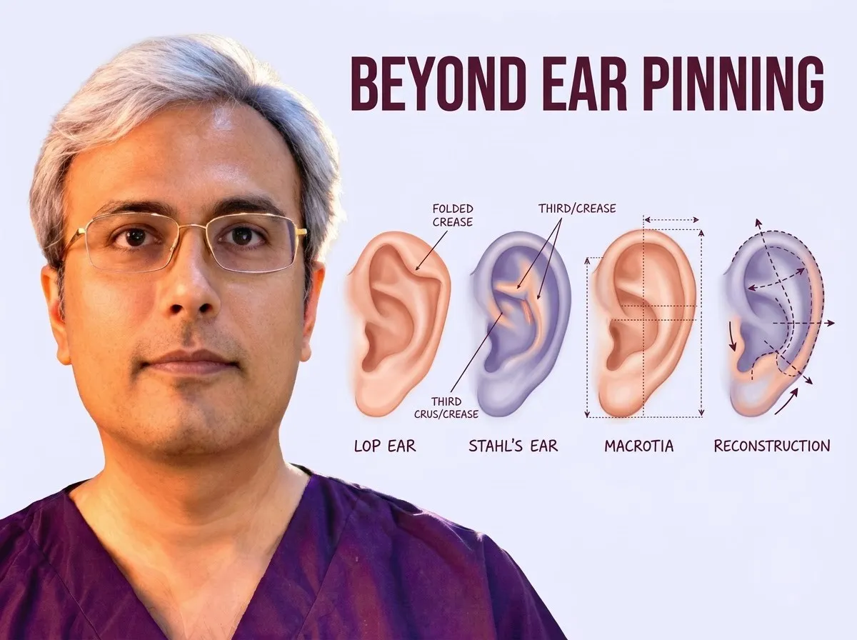
Ear differences range from simple outward protrusion to complex variations in cartilage shape and framework volume. When cartilage is folded tightly, compressed, or partially absent, standard setback sutures cannot restore balanced ear contours on their own.
Dr. Vishal Purohit, an M.Ch otoplasty surgeon in Jaipur, carefully evaluates structural cartilage deformities to determine whether specialized reshaping techniques are needed beyond conventional ear pinning.
Understanding why ears stick out helps clarify the boundary between standard ear pinning and procedures designed to release, trim, or rebuild abnormal cartilage structures.
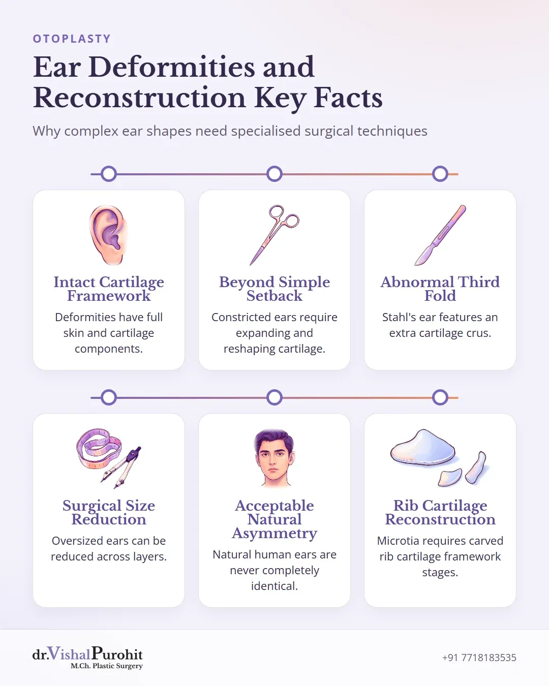
Prominence Is Only One Shape Difference
Ear differences are broadly divided into deformities and malformations12. Understanding this distinction helps clarify why standard ear pinning alone may not address every concern.
Deformations
Ear deformities feature an abnormal shape despite having fully developed skin and cartilage components1. The structural framework is intact13. These variations often result from mechanical forces inside or outside the uterus, such as pressure during development or abnormal muscle insertions13. Common examples include:
Dr. Purohit’s note: In his practice in Jaipur, Dr. Vishal Purohit accepts these presentations for surgical correction, assessing children from about 5 to 6 years of age. Newborn ear molding is not offered in his clinic.
Malformations
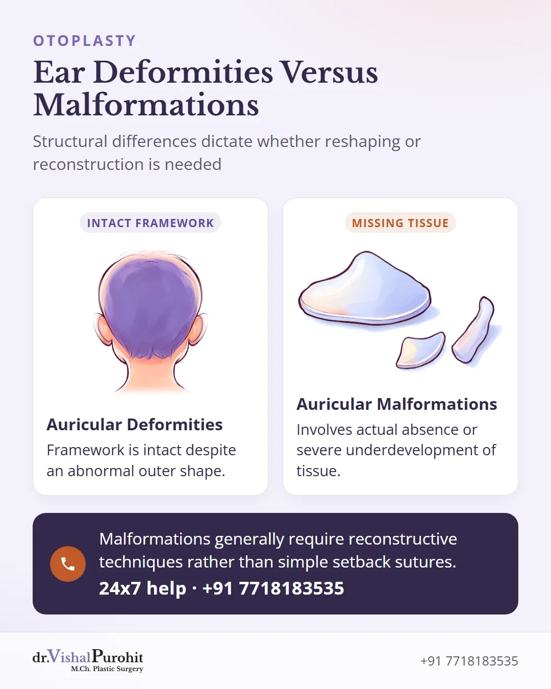
Auricular malformations develop from early embryologic disruptions, typically between the fifth and ninth gestational weeks, or genetic factors1. Unlike deformities, malformations involve an actual absence or severe underdevelopment of skin and cartilage1. Conditions in this category include microtia, anotia, cryptotia, and constricted ears1.
Because structural tissue is partially missing, malformations generally require reconstructive techniques rather than simple setback sutures14.
Lop and Cup Ears
A lop ear features an acute downward folding of the superior ear, involving the helix and scapha5. In contrast, a cup ear combines the downward folding of a lop ear with features of a prominent ear, presenting a deep, cup-shaped concha, a deficient upper helical rim and antihelical crus, and reduced vertical height5.
Key points
- Lop and cup ears are constricted ear deformities involving downward folding and variable cartilage deficiency.
- Standard ear pinning alone cannot restore vertical height or correct the collapsed cartilage framework.
- Treatment requires tailored surgical techniques such as cartilage release, flaps, or grafting before setback.
These deformities form part of the spectrum of constricted ears56. In mild cases, only the helical rim is folded, but more complex presentations involve both cartilage deficiency and tightening of the upper framework57.
Why Pinning Is Not Enough
Standard ear pinning addresses ear projection, but it cannot resolve the underlying structural collapse of a constricted ear. Proper correction requires expanding and reshaping the folded framework58:
- Simple set-back sutures cannot restore vertical height or open a tightly curled helical margin56.
- Reshaping the lidded helix, using cartilage release, repositioning, or local tissue flaps, must be completed before standard setback techniques are applied58.
- Severe cup deformities may require cartilage grafts to reconstruct the deficient framework5.
At his practice in Jaipur, Dr. Purohit treats lop and cup ears. Each ear requires an individualised surgical plan to release cartilage constriction, reconstruct the upper contours, and restore balanced proportions59.
Stahl’s Ear
Stahl’s ear is a congenital ear difference where an abnormal third fold of cartilage, known as a third crus, crosses the upper ear toward the outer rim1011. This extra fold flattens the natural curve of the rim, widens the upper groove, and often accompanies an underdeveloped or missing superior fold101112. In some presentations, it creates an angular, pointed contour at the top of the ear13.
Key points
- Stahl’s ear involves an abnormal third cartilage fold that flattens the upper rim and can create a pointed contour.
- Surgical approaches include cartilage scoring, excision, reshaping stitches, or grafting to rebuild the missing superior fold.
- Treatment in Jaipur focuses on correcting both the extra fold and any underdeveloped natural ear curves.
Surgical Correction in Literature
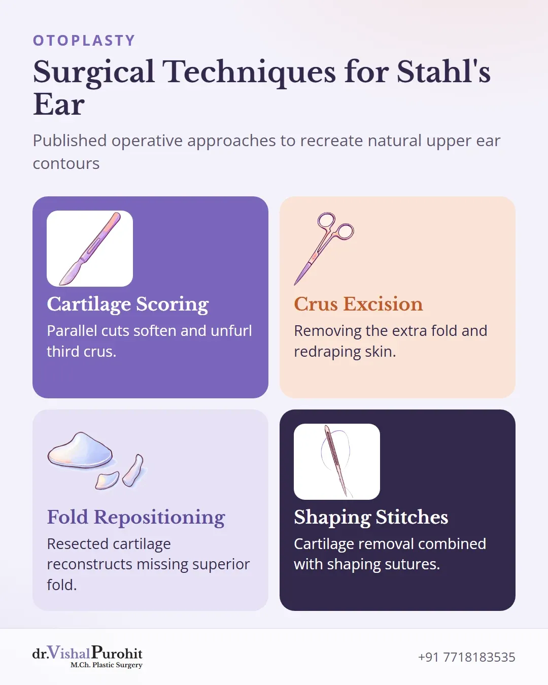
Because the anatomy varies, several operative approaches are described in published literature1214:
- Scoring the third crus with parallel cuts to soften and unfurl the cartilage fold15.
- Excision of the extra crus through an incision, redraping the overlying skin1516.
- Resecting the abnormal cartilage fold and repositioning or using it as a graft to reconstruct the missing superior fold101213.
- Combining cartilage removal with standard cartilage-shaping stitches to recreate a natural ear fold1517.
Dr. Vishal Purohit treats Stahl’s ear as part of his reconstructive and aesthetic ear surgery practice in Jaipur. Surgical correction addresses both the extra cartilage fold and any associated deficiency in the normal antihelical folds1215.
Large Ears and Uneven Ears
At his practice in Jaipur, Dr. Vishal Purohit treats large ears, uneven ears, and patients who need correction on one ear only.
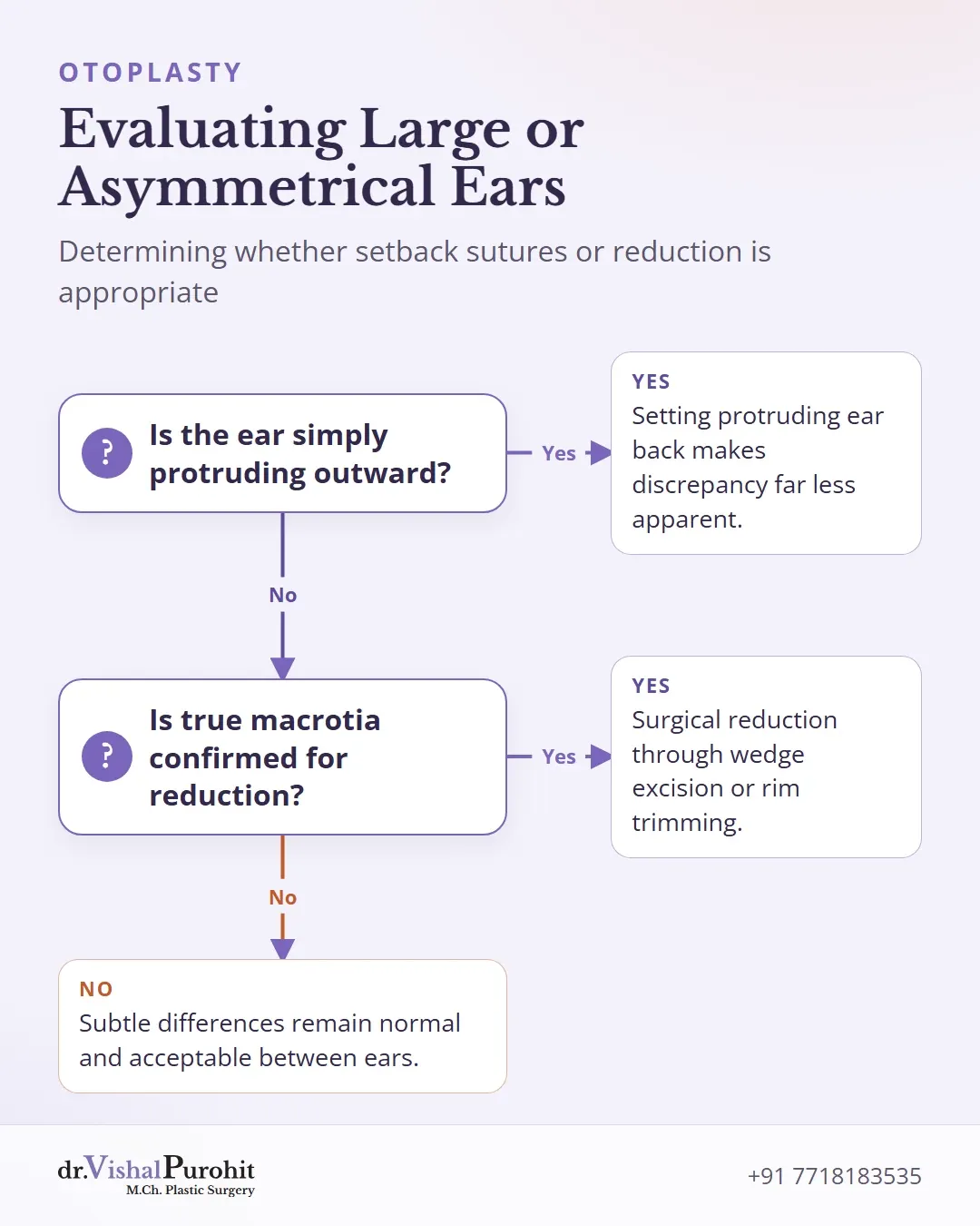
True macrotia, where an ear is genuinely oversized despite having normal proportions, is uncommon18. In many cases, an ear simply appears larger because it protrudes outward from the head, and setting the protruding ear back can make the size discrepancy far less apparent1819.
When surgical reduction of a large ear is required, operative approaches may include:
- Excising a wedge of tissue across all three layers of the auricle18.
- Reducing the helical rim by trimming a small rim segment alongside a crescent of scaphal cartilage20.
- Using chondrocutaneous advancement flaps along the helix to reshape and reduce the upper ear contour20.
For patients presenting with uneven ears or prominence on one side, surgery can address the affected ear to match the unoperated side1921. Natural human ears are never identical before or after surgery, and exact symmetry cannot be achieved. Subtle differences remain normal and acceptable because both ears are rarely viewed together at the same time22.
When Parts of the Ear Are Missing
Standard ear pinning cannot replace structural tissue when parts of the ear are underdeveloped or absent. Different conditions require specific reconstructive techniques rather than simple repositioning.
Microtia and Major Structural Absence
Microtia involves an underdeveloped external ear and requires formal surgical reconstruction rather than pinning23. The first stage of autologous ear reconstruction typically uses rib cartilage grafts carved into an auricular framework to rebuild the missing ear structure2324.
For a child with microtia, Dr. Purohit also arranges a hearing assessment with an ENT specialist.
Torn Earlobe Repair
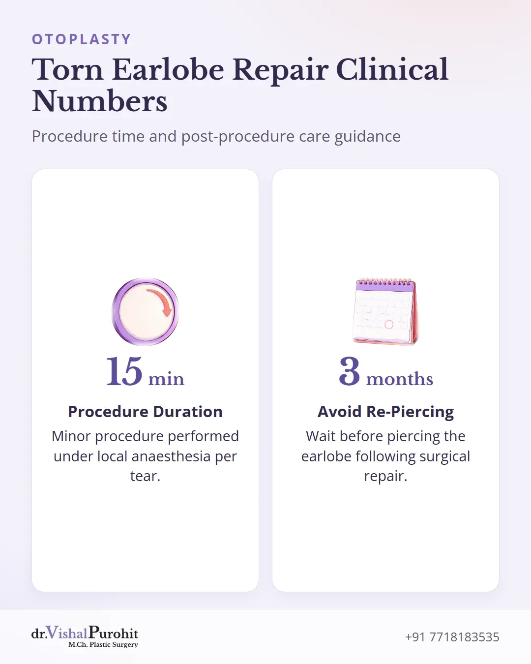
A torn earlobe is treated as a separate procedure and is not considered part of routine otoplasty23. The repair involves surgical excision of the tract or scar tissue followed by layered primary closure25. This minor procedure is typically performed under local anaesthesia and takes about 15 minutes per tear25. Dr. Purohit advises waiting 2 months before re-piercing the repaired earlobe; the details are on his earlobe repair page.
Consultation for Ear Surgery in Jaipur
Patients seeking correction for complex ear shapes, asymmetry, or prominent ears can explore options for otoplasty in Jaipur with M.Ch. plastic surgeon Dr. Vishal Purohit. Individualised operative planning ensures that each anatomical difference receives appropriate cartilage reshaping rather than routine setback sutures alone. For answers concerning recovery stages, dressings, and surgical details, read through the comprehensive otoplasty FAQs.
This is educational information, not medical advice.