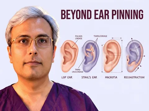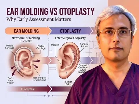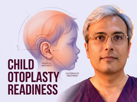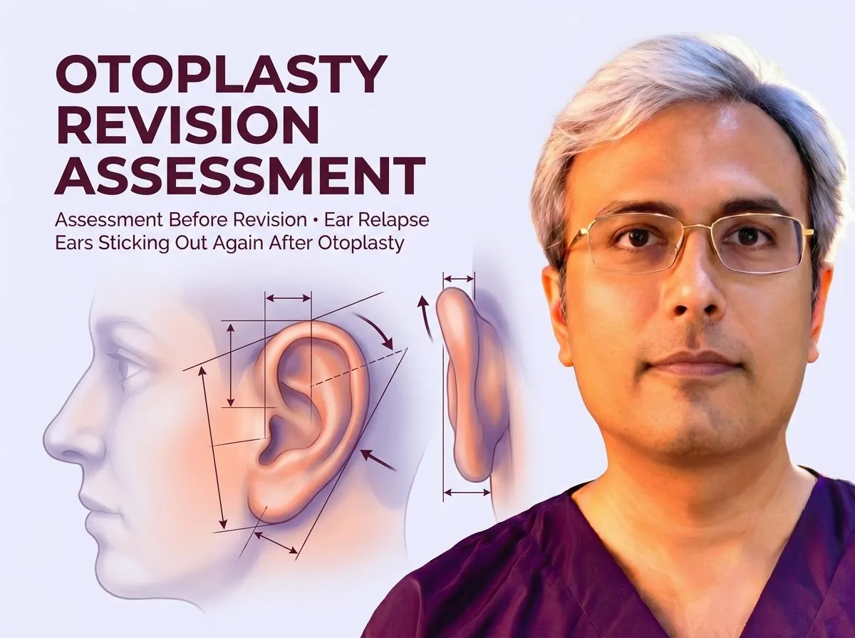
When an ear appears prominent after pinning surgery, tissue oedema or bandage pressure can easily cause confusion during the initial recovery phase. Learning how symmetrical ears should look after otoplasty helps families evaluate natural contours without mistaking normal healing for a failed surgical correction.
During follow-up consultations for otoplasty in Jaipur, Dr. Vishal Purohit assesses healing progression to distinguish temporary postoperative swelling from genuine positional changes requiring structural revision.
True structural relapse develops later and stems from mechanical tension, cartilage elasticity, stitch failure, or dressing displacement. A thorough surgical assessment evaluates remaining deformities and determines whether cartilage reshaping or suture adjustment is appropriate.
Early Swelling or True Recurrence?
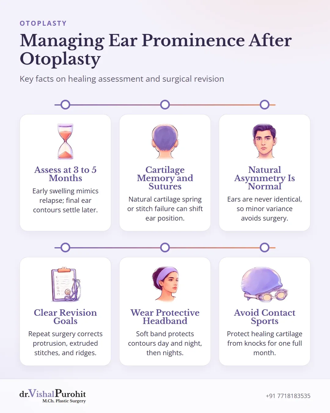
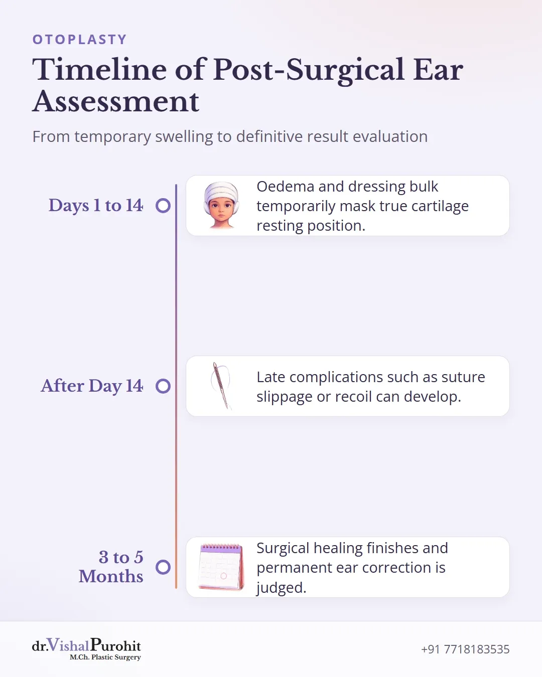
In the early postoperative phase, tissue oedema and the bulk of the surgical bandage can easily mislead both patients and families regarding ear position. Swelling alters local contours, while the pressure of the initial wrap temporarily masks the resting position of the cartilage.
Dr. Purohit’s note: In the protocol of Dr. Vishal Purohit, M.Ch. plastic surgeon in Jaipur, the final result is judged at 3 to 5 months.
Early changes do not necessarily represent a true surgical failure or loss of correction. In published clinical literature, otoplasty complications are categorized into early events occurring within the first 14 postoperative days and late events developing after 14 days1. Inadequate correction or loss of correction is reported in 6.5% to 12% of patients overall2. True late loss of correction can stem from factors such as suture slippage, suture extrusion, poor suture placement, thick or rigid cartilage, trauma, or noncompliance with postoperative dressings2. Furthermore, cartilage recoil or suture failure can cause a relapse over time3, with surgical relapse requiring intervention reported in approximately 10% to 30% of cases in craniofacial literature4.
Until surgical healing finishes and post-procedure swelling resolves completely, early ear prominence should not be mistaken for permanent structural recurrence.
Why Ears Can Move Forward Again
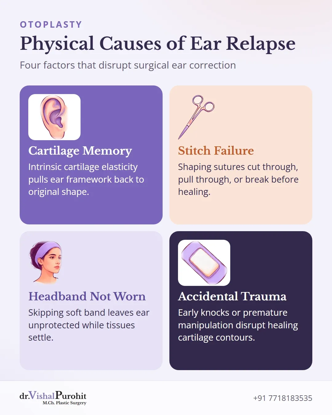
Recurrence is recognized as a frequent late complication following otoplasty5. Several physical and mechanical factors explain why an ear may gradually lose its correction:
Cartilage Memory
Ear cartilage possesses natural elasticity and intrinsic memory that pulls it back toward its original shape6. In stiffer or less pliable cartilage, higher tension is placed on the repair67. If sutures cannot hold this load until internal scar tissue matures and stabilizes the position, relapse can occur6.
Stitch Failure
Correction may be lost if internal shaping sutures cut through soft tissue or cartilage, pull through, or break before adequate healing occurs478.
Headband Compliance
At his practice, Dr. Vishal Purohit instructs patients to wear a soft headband day and night for 1 to 2 weeks after the surgical dressing is removed, followed by night-time wear for another 3 to 4 weeks to protect the repair while tissues settle.
- Trauma:
In general medical literature, surgical relapse or revision rates vary widely depending on technique and follow-up. Pooled analyses report recurrence rates around 3.2% and revision rates of 2.4%9, while other series note cartilage recurrence between 10% and 15%10, or relapse requiring intervention in 10% to 30% of cases4.
Other Results People Worry About
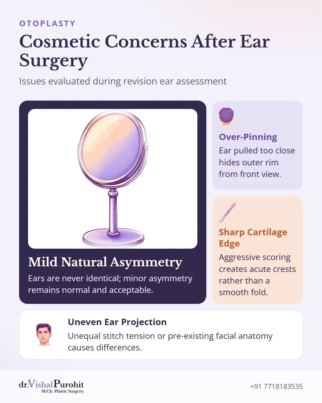
Beyond an ear sticking out again, patients seeking revision often worry about other cosmetic concerns from previous surgery:
Key points
- Previous ear surgery can result in over-pinning, sharp cartilage edges, or visible asymmetry.
- Mild asymmetry between the two ears is normal and acceptable because ears are naturally not identical.
- A revision assessment evaluates whether cartilage reshaping or suture adjustment is needed for significant concerns.
- Over-pinning: Overcorrection can pull an ear too close to the scalp, creating an unnatural, “pinned-back” look where the outer rim is hidden from the front view11.
- A sharp cartilage edge: Aggressive scoring, full-thickness cartilage incisions, or fracturing the cartilage can create visible ridges and acute crests along the fold of the antihelix rather than a smooth, natural curve111213.
- Unevenness between ears: Differences in position, projection, or shape can occur if suture tension is unequal or if underlying differences in ear size or facial anatomy were present before surgery1314.
In his practice, Dr. Vishal Purohit explains to patients that the two ears are never exactly alike before or after surgery, and perfect symmetry cannot be achieved. Because nobody sees both ears fully at the same time, mild natural asymmetry remains normal and acceptable. If minor asymmetry persists after healing, surgical correction may often be avoided13. When significant contour deformities or imbalances remain, an assessment for revision determines whether cartilage reshaping or suture adjustment is needed1415.
How a Revision Is Planned
Planning a revision begins with a detailed medical history and physical examination. In any discussion before repeat surgery, previous ear operations must be evaluated, as earlier interventions frequently leave scar tissue that can influence surgical planning and operative outcomes16.
Assessment and Photographs
Dr. Purohit evaluates revision cases, including patients who underwent their initial surgery elsewhere. A thorough clinical assessment identifies the nature of the residual deformity or recurrence.
Preoperative discussions clarify what secondary surgery can realistically achieve; this is acceptable because nobody sees both ears fully at the same time.
For a revision assessment, Dr. Purohit asks for photographs from the front, the back and both sides, with side views at 45, 90 and 135 degrees.
What Can Be Corrected
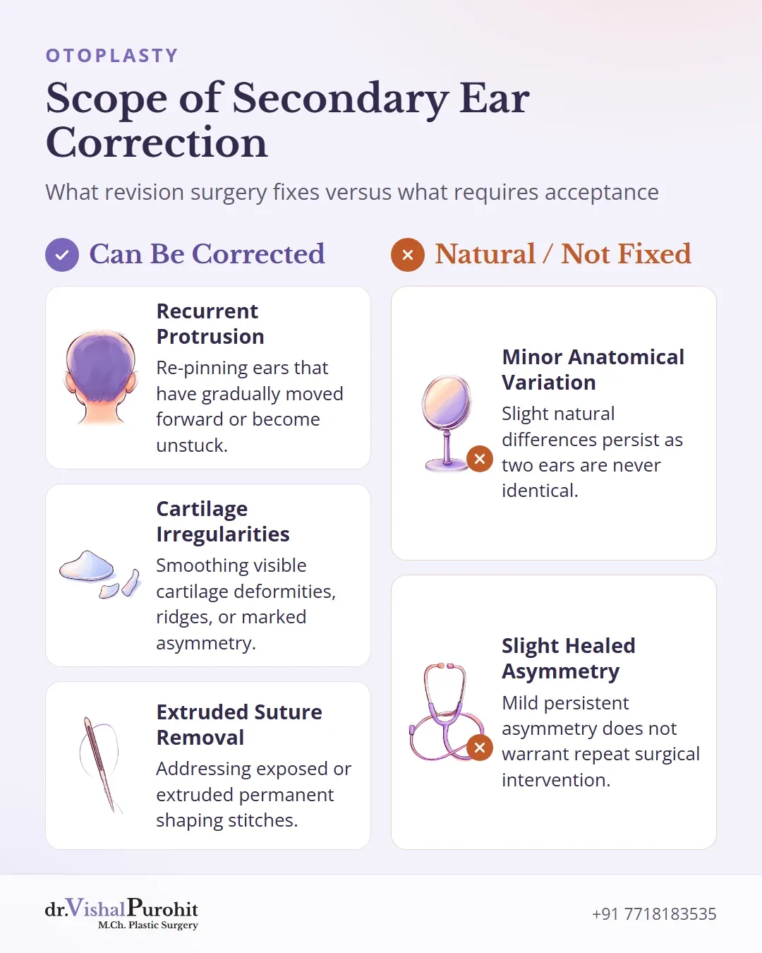
Revision surgery addresses visible issues remaining from previous procedures:
- Recurrence of protrusion or ears that have become unstuck1718
- Cartilage deformities, irregularities, or asymmetry1819
- Extruded permanent stitches17
Surgical correction combines cartilage-shaping stitches (Mustardé and Furnas type) with cartilage scoring or trimming, chosen for the specific ear. Any smaller adjustment is discussed at the review; a revision is charged if it needs general anaesthesia.
Protecting the Result
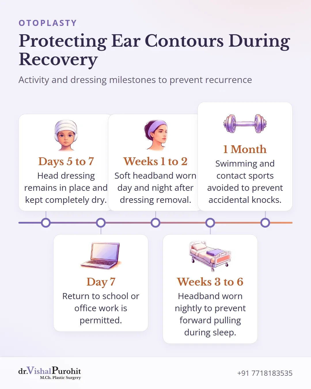
After otoplasty, protecting the ear position during healing is essential. In literature reviews, nocturnal headbands help prevent inadvertent forward pulling of the auricle while sleeping2021, and external pressures during rest can cause cartilage contours to buckle or shift22. Across multiple studies, pooled recurrence rates after otoplasty remain around 2.8%23.
In Dr. Vishal Purohit’s practice, the initial head dressing remains in place for 5 to 7 days and must be kept dry at all times, with the first dressing change scheduled on day 5. Once this head dressing is removed, patients transition to wearing a soft headband.
To avoid surgical site trauma and displacement:
- Return to school or office work is permitted at about 7 days.
- Swimming and contact sports such as cricket, football, or wrestling must be avoided until 1 month after surgery.
- Accidental knocks or manipulation of the healing cartilage must be prevented throughout early recovery.
Guidance regarding head dressings and safely resuming physical activity is discussed in dedicated posts on dressing care and return to sport.
Planning Revision Ear Surgery in Jaipur
Secondary ear procedures require careful evaluation of underlying scar tissue and cartilage contours. For individuals considering otoplasty in Jaipur, an in-person assessment clarifies achievable goals and operative techniques. Reviewing clinical details and otoplasty FAQs helps patients understand surgical timelines, healing phases, and postoperative protection.
This is educational information, not medical advice.