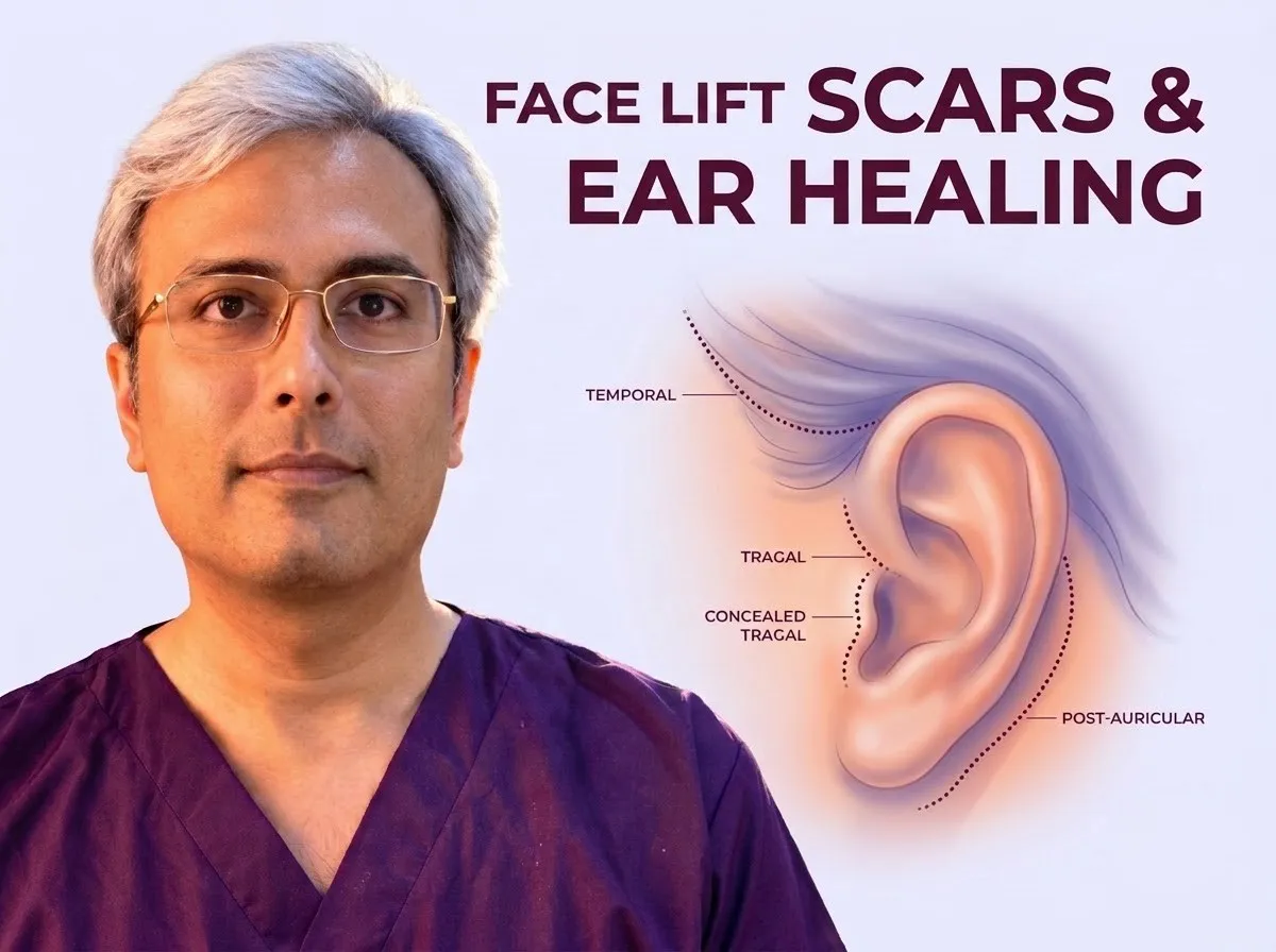
Facelift incisions are planned along natural anatomical borders and hair-bearing margins to blend into surrounding contours. The precise pattern depends on individual facial anatomy and the degree of correction required. Patients evaluating structural changes can also read about Mini, SMAS and Deep-Plane Facelift: What Do the Names Tell Patients? to understand underlying surgical techniques.
When evaluating incision placement and anatomical balance for facelift surgery in Jaipur, Dr. Vishal Purohit aligns surgical vectors with individual skin margins to protect hair-bearing borders and earlobe position.
Surgical cuts fade gradually over months, but they never completely disappear. Tension-free skin closure and deep tissue support help protect healing edges from widening. Careful preoperative planning preserves the natural sideburn position, prevents earlobe distortion, and supports balanced healing across different skin complexions.
Where Facelift Incisions Are Placed
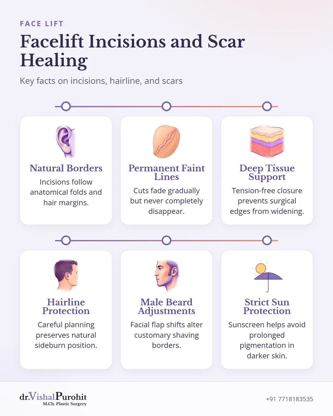
Facelift incisions are planned along natural anatomical borders and hair-bearing areas to blend into surrounding contours12. The exact route and length depend on patient anatomy and the extent of correction required13.
Hairline and Temporal Markings
In the upper face, incisions are placed either within the hair of the temple or along the anterior temporal hairline13.
- A post-hairline temporal incision sits concealed within the temporal scalp14. It is selected when minimal temporal skin is removed or when specific vectors of elevation allow the hairline to remain stable1.
- A pre-hairline incision runs along the margin of the temporal hair tuft or sideburn15. This position is considered when patients have short sideburns, in secondary facelift surgery, or when removing excess skin inside the hair would displace the hairline too far back from the corner of the eye16.
Incisions Around the Ear
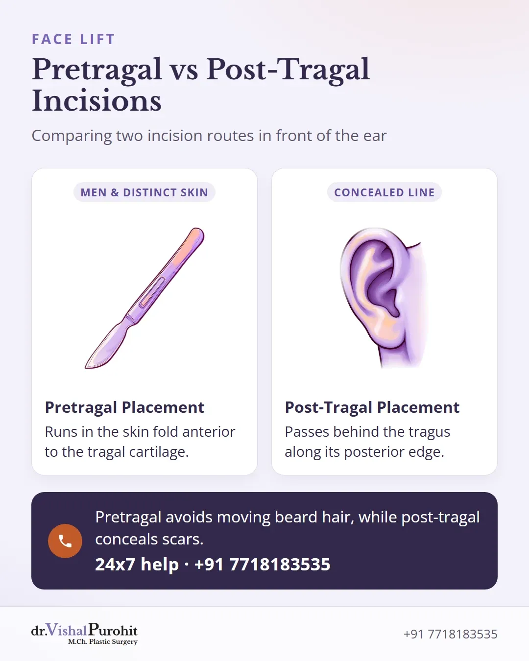
In front of the ear, the cut follows the curve of the helical rim downward16. Surgeons position this preauricular line either in front of or behind the tragus:
- Pretragal placement: The incision runs in the natural skin fold just anterior to the tragal cartilage15. This approach is often chosen in men to prevent moving beard hair onto the tragus, or when cheek skin differs markedly in appearance from tragal skin157.
- Post-tragal (retrotragal or intratragal) placement: The incision passes behind the tragus along its posterior edge to conceal the surgical line17. It requires careful contouring to avoid distortion of the tragus15.
Below the tragus, the marking curves around the earlobe attachment, preserving a natural boundary12.
Extent of the Lift and Incision Length
The posterior extension behind the ear depends on the degree of neck laxity3. In limited or short-scar approaches, the incision ends at the bottom of the earlobe or extends only minimally behind it358. When laxity in the neck and lower face is significant, the incision extends along the retroauricular groove and into the hairline of the posterior scalp to accommodate skin redraping137.
Dr. Purohit’s note: In his Jaipur practice, Dr. Purohit adapts surgical planning to each individual’s anatomical needs.
How Scars Mature and Fade
Surgical incisions fade over time, but they never completely disappear. Dr. Vishal Purohit explains to patients in Jaipur that while incisions are carefully placed along natural anatomical contours, scars are never invisible or undetectable.
The Maturation Timeline
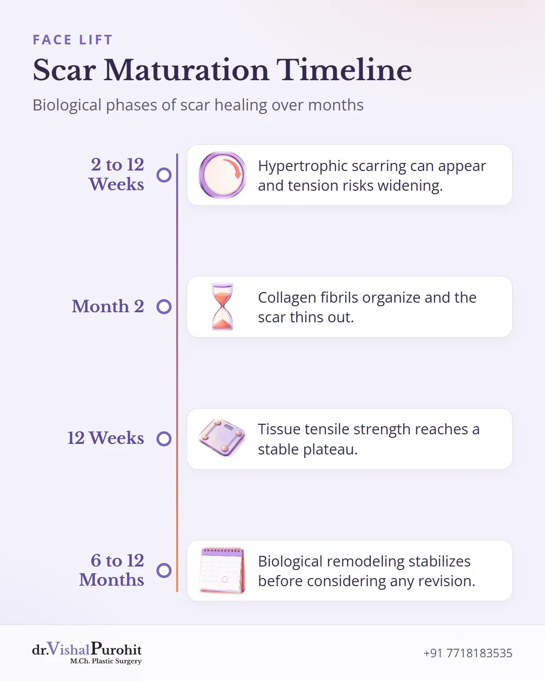
Healing is a gradual biological process that unfolds over many months:
Early Healing
In face lift surgery, hypertrophic scarring can appear as early as 2 weeks postoperatively and usually develops within the first 12 weeks9. Excessive wound tension or inadequate deep-layer support significantly increases the risk of widened or thickened scars910.
Collagen Remodeling
The formal scar maturation phase begins during the second month of wound healing, marked by a decrease in fibroblast density11. As collagen fibrils become more organized and lose water, the scar thins out11.
Long-Term Maturation
Tissue tensile strength rises progressively and reaches a plateau by 12 weeks, though it never regains the original strength of uninjured skin11. Scar maturation continues for years, and the final appearance of a surgical scar cannot be fully determined until a year after surgery11.
Because tissue remodeling takes months, secondary surgical procedures or excisions for hypertrophic scarring are delayed for at least 6 months to a full year to allow complete biological stabilization911.
Hairline Shift and Hair Loss Near the Incisions
Incisions for a facelift must be planned around the sideburns and temporal hair to maintain natural anatomical boundaries. Placing an incision deep inside the temporal hair can pull non-hair-bearing cheek skin upward and backward1213. When skin redundancy is large or temporal hair is sparse, this shift can lead to backward distortion of the temporal hairline and elevation or loss of the sideburn121314. In literature, advancing facial skin flaps into hair-bearing scalp without accounting for tissue movement creates an operated appearance1315.
Key points
- Incisions must be planned around sideburns and temporal hair to preserve natural boundaries.
- Deep temporal incisions can pull cheek skin upward, distorting the hairline and elevating or losing the sideburn.
- Hair loss along incision lines is minimized by gentle tissue handling and low closure tension.
Hairline-Preserving Incision Approaches
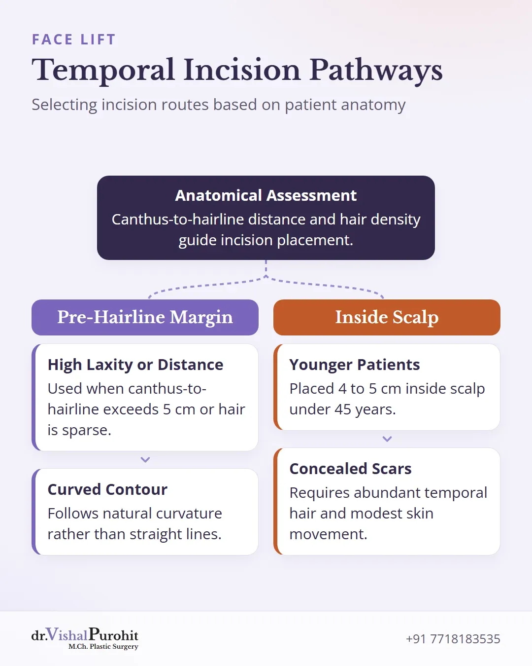
To prevent sideburn and hairline displacement, surgical literature outlines specific incision choices based on individual anatomy1314:
- An incision positioned directly along the temporal hairline is selected when the lateral canthus-to-hairline distance exceeds 5 cm, when marked skin elevation is anticipated, or when sparse temporal hair cannot tolerate upward movement1315.
- Traditional incisions placed 4 to 5 cm within the temporal scalp are typically reserved for patients under 45 years of age with abundant hair and modest predicted skin shifts14.
- Incisions along the hairline should follow its natural curvature rather than a straight line to prevent noticeable, distorted scar lines1216.
Incisional Hair Loss
Localized hair loss along incision lines, known as scar or cicatricial alopecia, can occur after surgery1718. Surgical evidence notes that hair loss is avoided by minimizing closure tension and using gentle tissue handling18. Overuse of electrocautery, tight sutures, and staples damages hair roots18. For patients with noticeable incisional hair loss or displaced sideburns, hair transplant procedures can reconstruct a natural hairline pattern1217. Dr. Vishal Purohit plans surgical incisions with anatomical precision during consultations in Jaipur.
The Earlobe and Pixie Ear
A pixie ear deformity is an unnatural, elongated, or pulled-down appearance of the earlobe following a facelift1920. It develops when excessive tension is placed across the lower flap and cheek skin during closure, or when postoperative wound contracture pulls down on an otherwise well-positioned lobe1921. Most acquired cases of this deformity arise after rhytidectomy22.
Key points
- Pixie ear deformity is a pulled-down earlobe after facelift surgery.
- It results from excessive tension or wound contracture during healing.
- Surgical revision techniques can correct the deformity.
Prevention During Surgery
In his practice in Jaipur, Dr. Vishal Purohit performs SMAS plication and suspension facelifts, anchoring underlying support to deeper facial tissues rather than letting the superficial skin carry tension.
In the surgical literature, technical steps to prevent pixie ear include:
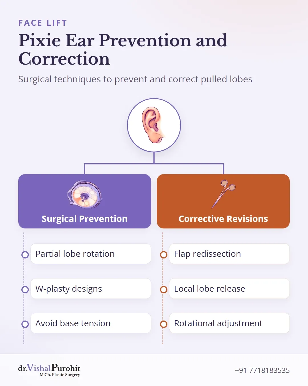
- Partial rotation of the lower earlobe, which has been documented as a reliable method to prevent deformities during face lift surgery2023.
- Incision designs such as W-plasty around the incision site2022.
- Avoiding excess tension at the base of the earlobe to prevent secondary traction during healing1921.
Corrective Approaches
If a pixie ear deformity develops, several surgical revision techniques exist in published plastic surgery literature1921:
- Complete flap redissection: Reopening the previous facelift incisions, freeing the flaps, excising excess skin, and repositioning the flap and earlobe superiorly192124. This comprehensive approach generally requires waiting until sufficient skin laxity develops192124.
- Local lobe release: Incising around the base of the earlobe, which releases the scar and allows the lobe to retract upward192124. The resulting trough is closed primarily, trading earlobe position for a small scar below the lobe2124.
- Rotational adjustment: Performing a partial earlobe rotation to restore normal lower curvature and volume2023.
Men: Beard Skin and Shaving Changes
In male facelift surgery, the location and pattern of facial hair require specific planning2526. Repositioning facial skin pulls hair-bearing beard tissue toward the ear, which can alter normal shaving boundaries2627.
When cheek and neck skin flaps are mobilized, hair follicles may shift closer to the ear margin or directly onto the tragus2627. Surgeons also take care not to pull hair-bearing neck skin onto the ear during redraping26.
Follicle Management and Postoperative Shaving
To manage hair-bearing skin around the ear, published surgical techniques describe several steps:
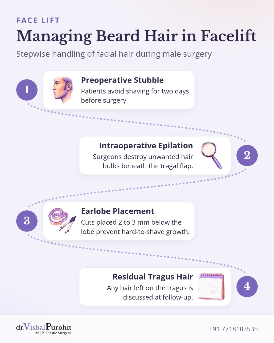
- Preoperative stubble: Patients may be instructed not to shave for two days before surgery so follicles project clearly beneath the flap2628.
- Intraoperative epilation: Surgeons can trim subcutaneous fat and destroy or cauterize unwanted hair bulbs beneath the tragal flap under magnification272829.
- Earlobe placement: Incisions placed 2 to 3 mm below the earlobe junction help prevent beard growth in regions that are difficult to shave29.
Because beard hair grows in cycles, follicle shift cannot always be fully removed during the operation27. If bearded skin remains on the tragus, literature suggests waiting 4 to 6 months before considering secondary electrolysis27.
Darker Skin, Pigmentation and Scar Care
In deeper or olive complexions common among Indian patients, pigmented skin inherently heals with post-inflammatory hyperpigmentation (PIH)30. Increased pigmentation of scars represents mostly a post-inflammatory response that can take several weeks or 3 to 18 months to fade3031. This discoloration corresponds to the area of tissue inflammation31.
Sun Protection and Consultation Review
Strict sun protection and sun avoidance are essential to avoid aggravating hyperpigmentation or delaying pigment resolution30313233. Literature recommendations include regular application of broad-spectrum sunscreen with a minimum SPF of 30, such as formulations containing zinc oxide or titanium dioxide3233.
A personal or family tendency toward raised scars requires direct discussion prior to surgery:
- Keloids show a predilection for dark-skinned individuals and young persons34.
- Wounds closed under excessive tension carry an increased risk of hypertrophic or keloid scarring34.
- The risk of developing abnormal scars is assessed during the initial consultation with Dr. Vishal Purohit in Jaipur.
Silicone and Scar Care
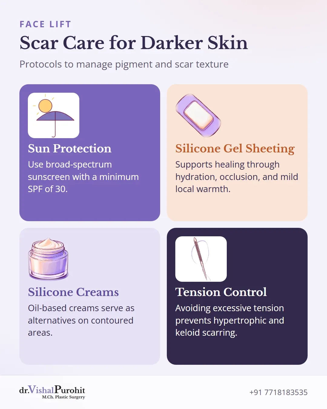
General surgical literature supports topical silicone for hypertrophic scar and keloid management35. Silicone gel sheeting has several randomized controlled trials supporting its safe use, working through hydration, occlusion, and mild local temperature elevation35. When sheeting is not feasible on contoured areas of the face or neck, silicone oil-based creams serve as an alternative35.
Dr. Vishal Purohit does not offer laser or skin resurfacing procedures. Postoperative scar-care protocols are individualized, and any persistent concerns regarding scar texture or pigmentation are addressed directly with the surgical team during clinical follow-up reviews.
Individualized Surgical Planning in Jaipur
Anatomical precision and delicate tissue handling ensure incisions heal as inconspicuously as possible. Patients seeking balanced facial rejuvenation can explore facelift surgery in Jaipur to review suitable surgical approaches. For additional questions regarding healing, incision placement, and tissue remodeling, browse our facelift FAQs to prepare for your clinical consultation.
This is educational information, not medical advice.