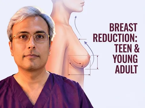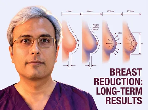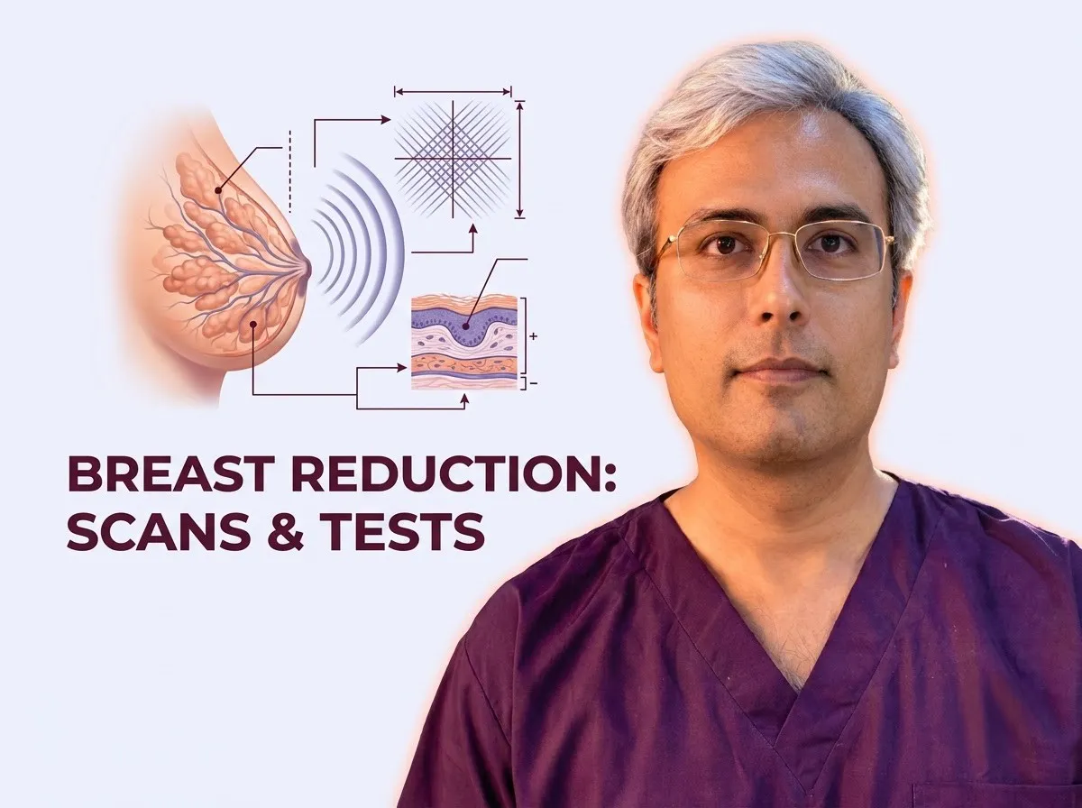
Evaluating breast health before a reduction mammaplasty helps surgeons identify unsuspected tissue alterations and establish an accurate baseline for subsequent imaging. In Dr. Vishal Purohit’s practice, a mammogram or breast ultrasound is required before surgery for every patient, with genetic counselling (BRCA1/BRCA2) arranged when family risk is high.
During a Breast Reduction Consultation: What to Expect, the surgical team reviews past medical concerns and assesses the breasts thoroughly. This structured evaluation helps ensure safe planning before altering the breast tissues.
Breast Checks at a Glance
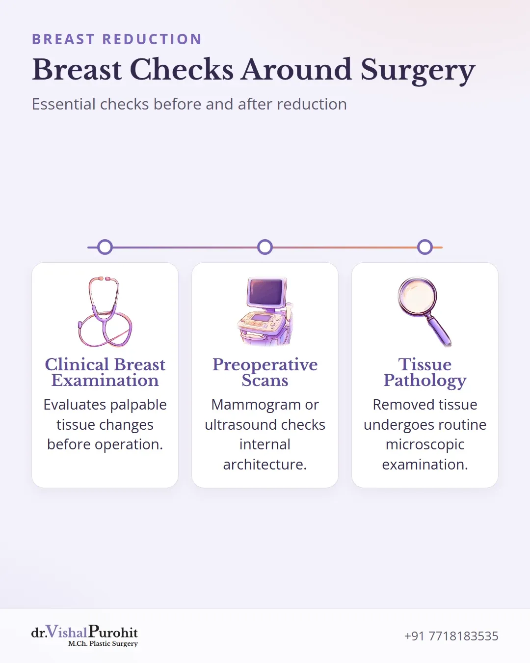
Before and after reduction mammaplasty, breast evaluation helps detect unsuspected tissue changes and establishes an accurate baseline. Published protocols describe the role of each assessment:
| Check | Who usually needs it | Why |
|---|---|---|
| Clinical breast examination | Patients undergoing reduction mammaplasty [1] | Evaluates the breasts during outpatient visits and on the day of surgery to identify palpable changes [1]. |
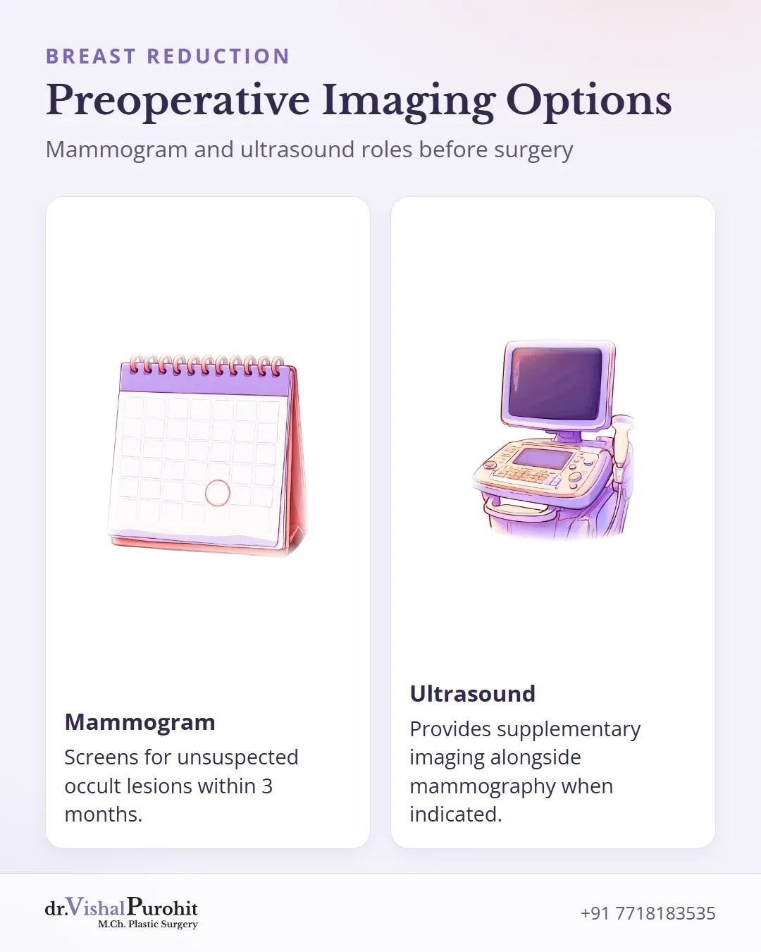
| Mammogram | Patients evaluated before surgery based on clinical protocol rather than a fixed age [1] | Screens for unsuspected occult lesions prior to surgery, typically performed within 3 months of the operation [1]. |
| Ultrasound | Patients where indicated by the reporting radiologist [1] | Provides supplementary imaging alongside mammography when further evaluation is needed [1, 2]. |
| Pathology of removed tissue | All reduction mammaplasty specimens [1, 3] | Histological examination identifies incidental proliferative changes or occult disease [1, 3]. |
| Future screening | Patients who have undergone breast reduction [4] | Continues long-term breast health monitoring, as reduction mammaplasty does not replace routine breast surveillance [4]. |
In his practice at Kalpana Aesthetics in Jaipur, Dr. Vishal Purohit requires a mammogram or breast ultrasound before surgery for every patient, arranging genetic counselling (BRCA1/BRCA2) when family risk is high.
All removed breast tissue is routinely sent for histopathology, and the result is provided to the patient as a printed report.
The Breast Examination at Consultation
Before planning a reduction mammaplasty, a comprehensive clinical evaluation is the essential first step. At Kalpana Aesthetics in Jaipur, consultations with M.Ch. plastic surgeon Dr. Vishal Purohit prioritize patient safety and an evidence-grounded assessment.
In Dr. Purohit’s practice, a mammogram or breast ultrasound is required before surgery for every patient, and genetic counselling (BRCA1/BRCA2) is arranged when family risk is high.
A thorough history and physical examination establish baseline breast health before any surgical intervention is undertaken. Key discussions during preoperative consultations include assessing:
- Personal expectations
- Surgical benefits
- Potential risks
- Family history of breast cancer [5]
Identifying personal or familial cancer history is critical. Patients with prior oncologic diagnoses or breast cancer history have demonstrated higher incidences of high-risk proliferative lesions compared to those presenting solely for symptomatic macromastia [1, 6].
Assessment of New Findings
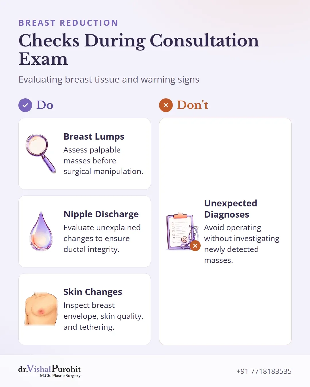
During the physical examination, the breasts and surrounding tissues are carefully evaluated. Any newly detected finding requires full clinical assessment prior to surgery:
- Breast Lumps: Palpable masses or lesions must be assessed prior to surgical manipulation [2]. Benign conditions such as fibrocystic changes, fibroadenomas, or adenosis may occur, but high-risk lesions or unexpected malignancies must be ruled out [3, 6].
- Nipple Discharge or Changes: Any unexplained nipple alterations must be evaluated to ensure ductal integrity and rule out underlying disease.
- Skin Changes: The breast envelope, skin quality, and induration or tethering are inspected [7].
Preoperative protocols aim to avoid unexpected diagnoses during standard reduction mammaplasty. This protects oncological safety and spares patients unnecessary secondary procedures [1].
Mammogram or Ultrasound Before Surgery?
Pre-operative breast evaluation before a reduction mammaplasty is not governed by a single blanket testing panel.
Instead, recommendations for breast imaging prior to surgery are individualized based on patient age, clinical symptoms, and individual risk profiles.
Age and Clinical Evaluation
The need for pre-operative imaging varies across different life stages:
Younger Women
Younger patients seeking breast reduction undergo careful clinical assessment [8], with decisions regarding imaging tailored to their specific presentation rather than routine screening mandates.
Older Women
Breast reduction in older women involves addressing distinct age-related expectations and surgical considerations [9], where background breast tissue changes and screening history play an important role in surgical planning.
After Surgery
Some surgical series report yearly breast imaging after a reduction [10].
Tailored Assessment Over Universal Panels
Published medical literature emphasizes that breast reduction candidates should be evaluated on an individual basis rather than subjected to an arbitrary panel of tests [9]. Routine breast imaging is not applied identically to every individual, as risk factors, family history, and symptoms guide the selective use of modalities such as mammography or ultrasonography.
Dr. Purohit’s note: In Dr. Purohit’s practice at Kalpana Aesthetics in Jaipur, a mammogram or breast ultrasound is required before surgery for every patient, and genetic counselling (BRCA1/BRCA2) is arranged when family risk is high.
Testing the Removed Tissue
During breast reduction surgery (reduction mammaplasty), excess glandular tissue, fat, and skin are excised to relieve physical symptoms and reshape the breast.
Key points
- All tissue removed during breast reduction is sent for microscopic pathological evaluation.
- Most reduction specimens show normal tissue or benign changes such as fibrocystic disease.
- Routine histopathology ensures that any incidental or occult proliferative findings are identified.
Pathological evaluation of the resected tissue allows for microscopic examination of the breast parenchyma. In Dr. Vishal Purohit’s practice at Kalpana Aesthetics in Jaipur, all removed breast tissue is always sent for histopathology, and the result is provided to the patient as a printed report.
Pathological Findings in Reduction Specimens
In published literature discussing proliferative breast lesions, the World Health Organization defines these changes as cytologically and architecturally diverse proliferations confined to the mammary gland. These changes originate primarily from the terminal ductal-lobular unit and less often from lactiferous ducts [11].
Studies identify several benign and atypical proliferative findings that can appear within breast tissue [11]:
- Usual ductal hyperplasia (UDH) [11]
- Flat epithelial atypia (FEA) [11]
- Atypical ductal hyperplasia (ADH) [11]
- Atypical lobular hyperplasia (ALH) [11]
- Pseudoangiomatous stromal hyperplasia (PASH) of mesenchymal origin [11]
Because incidental or occult proliferative changes may be discovered within tissue removed during mammaplasty, microscopic evaluation can identify these lesions [11].
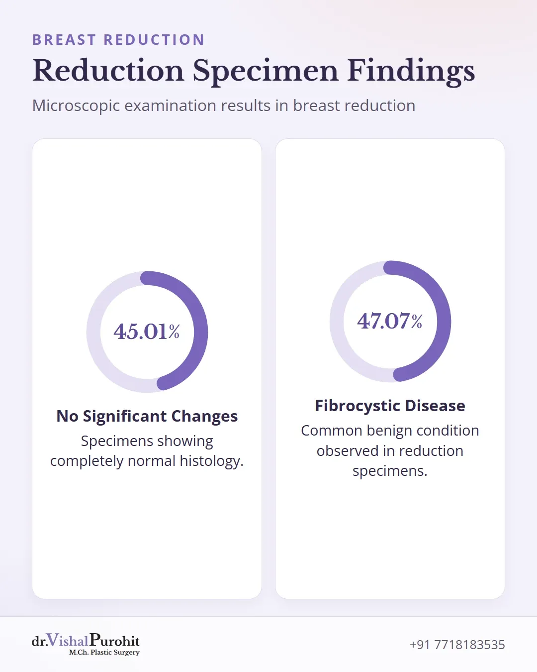
In one study of breast reduction specimens, 45.01% showed no significant changes and 47.07% showed fibrocystic disease, a benign condition; smaller numbers showed adenosis or fibroadenoma [3]. Studies comparing groups of patients report more high-risk lesions in women who have had breast cancer than in women having a reduction for large breasts alone [6].
Breast Screening After a Reduction
Changes on Later Breast Imaging
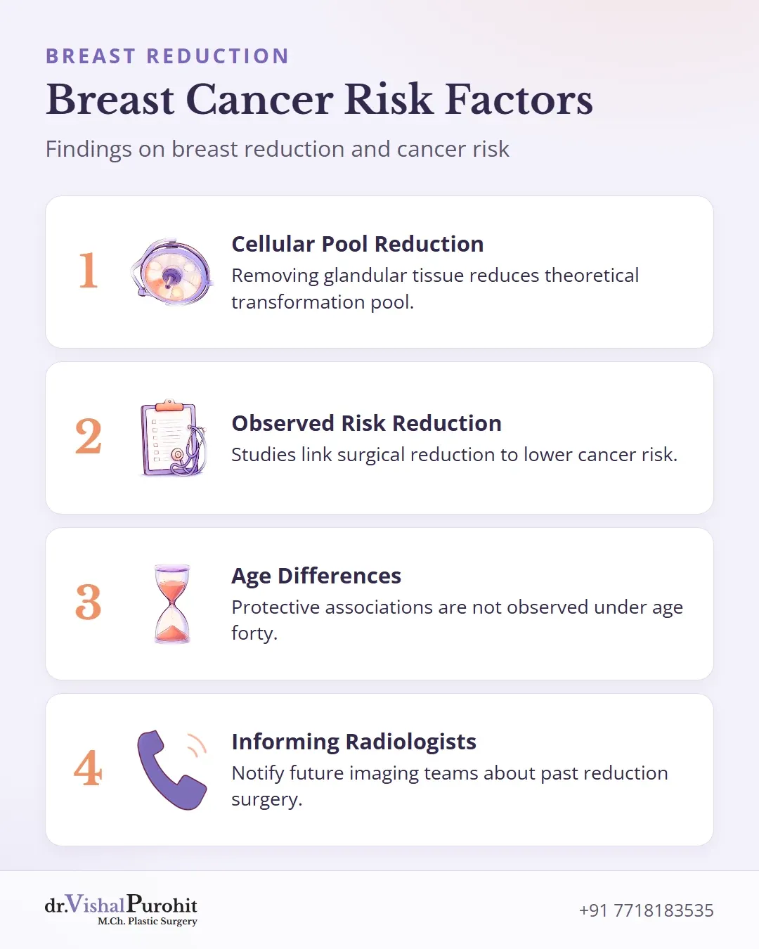
Because surgical alteration changes the internal architecture of the breast, informing the radiologist and imaging team about past reduction surgery is essential when attending future breast screenings.
What Studies Report on Breast Cancer Risk
Clinical research has evaluated whether removing breast tissue influences subsequent cancer rates:
- Tissue mass and cellular risk: Having a larger volume of glandular tissue intuitively increases the number of cells that could theoretically undergo malignant transformation [12]. Glandular tissue reduction is thought to lower this cellular pool [12].
- Observed risk reductions: Several studies report that surgery to reduce breast size lowers breast cancer risk later in life [12]. For instance, a nationwide study in Denmark noted that surgical reduction in breast size was associated with a 50 percent reduction in subsequent breast cancer risk, mirroring a 39 percent reduction reported in an earlier Danish series [12]. In a United States population-based study, a relative risk of 0.7 was observed, though based on small patient numbers and without statistical significance [12].
- Age differences: These protective associations are not uniform across all groups. In the Danish cohort, a reduction in risk was not observed among women under the age of 40 at the time of surgery [12].
Breast Health and Reduction Surgery in Jaipur
Undergoing thorough breast evaluation before and after surgery ensures that underlying tissue characteristics are properly managed. In Dr. Vishal Purohit’s practice, a mammogram or breast ultrasound is required before surgery for every patient considering breast reduction surgery in Jaipur, with genetic counselling (BRCA1/BRCA2) arranged when family risk is high.
All removed breast tissue is sent for histopathology, and the result is provided to the patient as a printed report alongside clear guidance on breast reduction and cancer screening.
This is educational information, not medical advice.