
Periorbital darkness is often mistaken for a single condition, but shadows arise from several distinct anatomical causes. Structural depressions along the infraorbital rim may benefit from hyaluronic acid dermal filler, whereas skin pigmentation and vascular congestion remain unaffected by injectable volume.
Lower eyelid anatomy requires careful clinical assessment because fluid retention or tissue laxity can alter treatment outcomes. Understanding Under-Eye Fillers vs Lower Eyelid Surgery: How Are They Different? helps clarify when non-surgical volume is appropriate and when structural fat herniation requires a surgical approach.
Hollowing Pigment and Structural Eye Bags
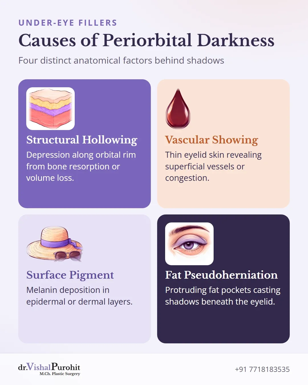
Periorbital darkness is a descriptive presentation rather than a single condition1. True dark circles arise from distinct anatomical factors1:
- Structural hollowing: a depression along the infraorbital rim caused by inherited anatomy, bone resorption, or tissue volume loss12.
- Vascular showing: thin eyelid skin revealing superficial vessels or congestion1.
- Surface pigment: epidermal or dermal melanin deposition1.
- Orbital fat pseudoherniation: protruding fat pockets creating a shadow below the lower eyelid12.
Hyaluronic acid dermal filler adds physical volume beneath a sunken groove along the orbital rim13. However, injectable filler cannot address every under-eye concern. It does not bleach superficial or constitutional melanin pigmentation1. It also cannot relieve vascular congestion or tighten lax tissue over herniated orbital fat pockets12.
Dr. Purohit’s note: Dr. Vishal Purohit assesses each person’s lower eyelid anatomy to identify whether a sunken hollow, surface pigment, or protruding fat dominates.
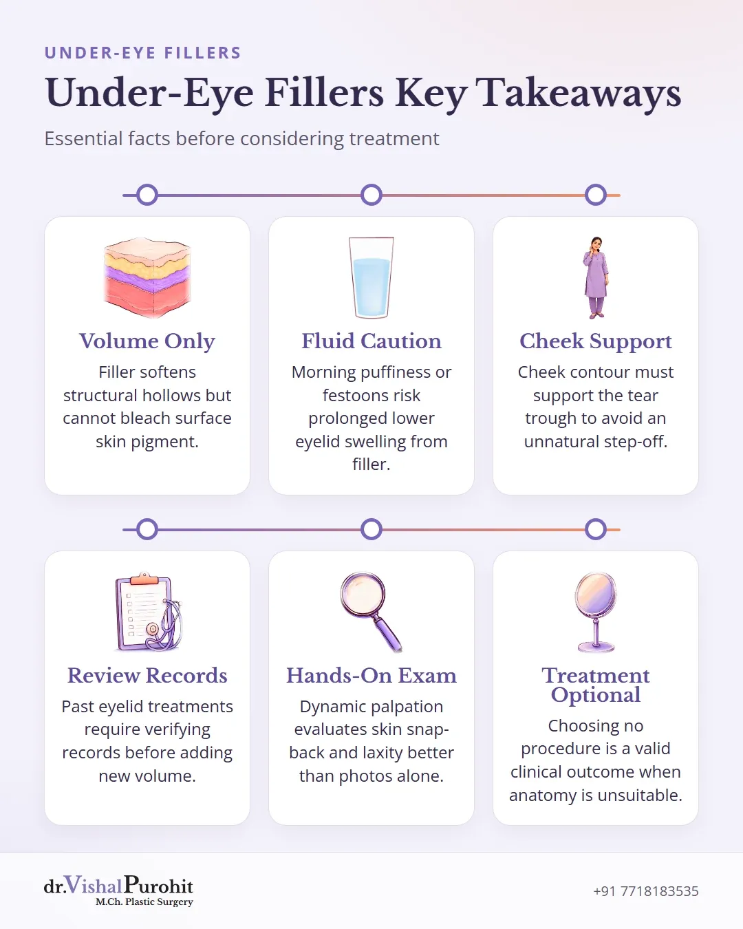
Filler is generally considered only when a structural volume deficiency causes the shadow. Choosing no treatment remains a valid clinical outcome, and cosmetic procedures are never mandatory.
Who May Be Unsuitable for Tear Trough Filler
Not every person seeking under-eye rejuvenation is a candidate for hyaluronic acid dermal filler. Certain anatomical presentations and physiological tendencies make treatment inadvisable.
Key points
- Hyaluronic acid dermal fillers are inadvisable for individuals with morning eyelid fluid retention, malar mounds, or festoons.
- Because hyaluronic acid attracts water, it can aggravate compromised lymphatic drainage and cause lasting swelling.
- Systemic health conditions must be evaluated and ruled out before considering under-eye cosmetic procedures.
Fluid Retention and Structural Changes
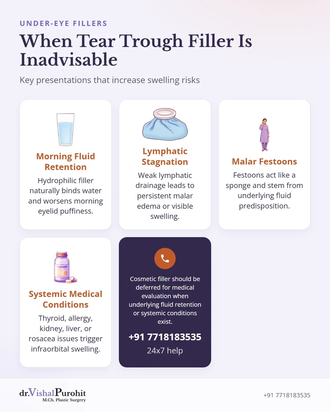
Patients presenting with morning eyelid fluid retention, malar mounds, or true festoons are generally poor candidates for tear trough filler45.
Hyaluronic acid naturally attracts and binds water to create volume56. In the delicate infraorbital area, this hydrophilic property can exacerbate fluid retention and worsen lymphatic stagnation45. In individuals with compromised lymphatic drainage, filler placement increases the risk of persistent malar edema or visible swelling45. Festoons themselves may act like a sponge and often stem from underlying lymphatic predisposition7.
Underlying Medical Assessment
Infraorbital swelling and malar edema may also be triggered by systemic conditions, such as allergy, thyroid eye disease, renal disease, liver disease, or inflammatory disorders like rosacea47.
Deferring cosmetic filler for medical evaluation avoids compounding baseline swelling47. At his practice in Jaipur, Dr. Vishal Purohit assesses candidates individually to determine suitability for non-surgical treatments. A medical evaluation helps rule out underlying causes before any cosmetic procedure is considered.
Why the Lid Cheek Transition Is Examined
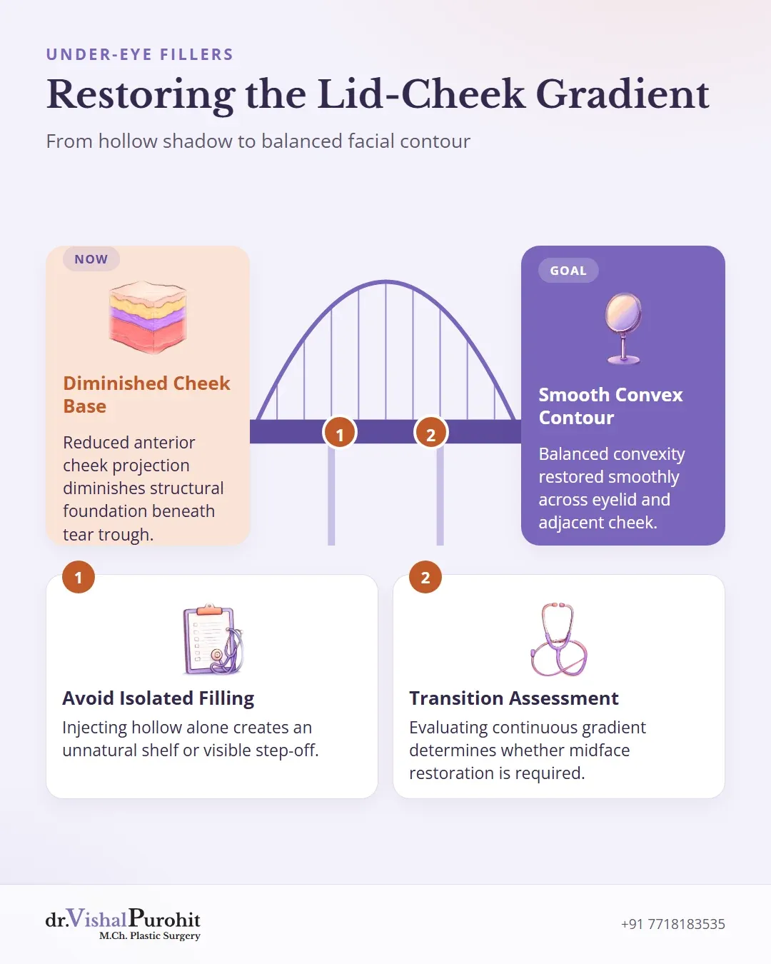
The lower eyelid and the anterior cheek form a continuous anatomic gradient that must be evaluated together89. Volume loss in the cheek can cause an elongated lid-cheek junction and accentuate under-eye hollows910.
A desirable cheek contour is convex rather than flat11. When anterior cheek projection is reduced, the structural foundation beneath the tear trough is diminished811. Injecting dermal filler into the under-eye hollow alone without cheek support can leave an exaggerated step-off or shadow beneath the treated area8. This can create the appearance of an unnatural shelf or overfilled lower eyelid, because smooth convexity from the eyelid across the midface has not been restored8.
Dr. Vishal Purohit assesses the relationship between the lower lid and the midface during consultation89. Whether a patient requires isolated under-eye treatment, midface volume restoration, or another approach is not confirmed in advance and is decided at the consultation. Evaluating this transition helps ensure that contouring remains balanced across adjacent facial structures89.
Clinical History and Prior Eyelid Interventions
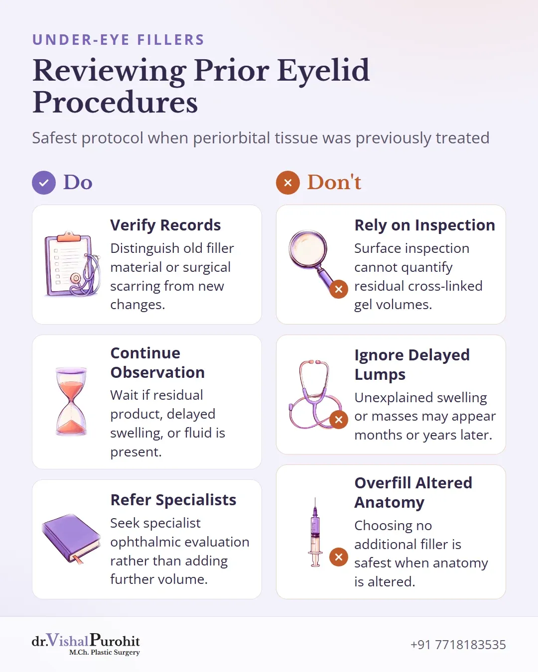
Patients who have had earlier cosmetic injections or eyelid surgery require a detailed records check before planning any further treatment1213. A history of previous facial procedures substantially guides diagnostic evaluation and changes treatment decisions12. In his practice, Dr. Vishal Purohit assesses past interventions thoroughly during consultation.
Key points
- Prior cosmetic procedures significantly alter anatomy and affect future treatment decisions.
- Physical examination alone cannot confirm remaining filler volume or identify specific past materials.
- Choosing observation or review is often safer than adding more filler.
Physical examination alone is not sufficient to assess tissue altered by prior procedures413. Surface inspection cannot identify the specific types of residual cross-linked gel or determine the exact remaining volumes retained beneath delicate eyelid skin14. Unexplained swelling, delayed lumps, or orbital masses may appear months or years after an initial procedure1215.
A complete reassessment helps determine the safest course of action:
- Verifying previous records to distinguish old filler material or surgical scarring from new changes1213.
- Continuing observation if residual product, delayed swelling, or fluid retention is already present41215.
- Referring for specialist ophthalmic evaluation or elective review rather than adding further volume1213.
Choosing no additional filler is often appropriate when earlier procedures have already altered periorbital anatomy1216.
Realistic Limits and Clinical Eyelid Assessment
A thorough in-person eyelid assessment evaluates skin snap-back, septal integrity, and true negative vector support. Photographs taken under flat lighting cannot replace dynamic physical palpation when ruling out swelling risks.
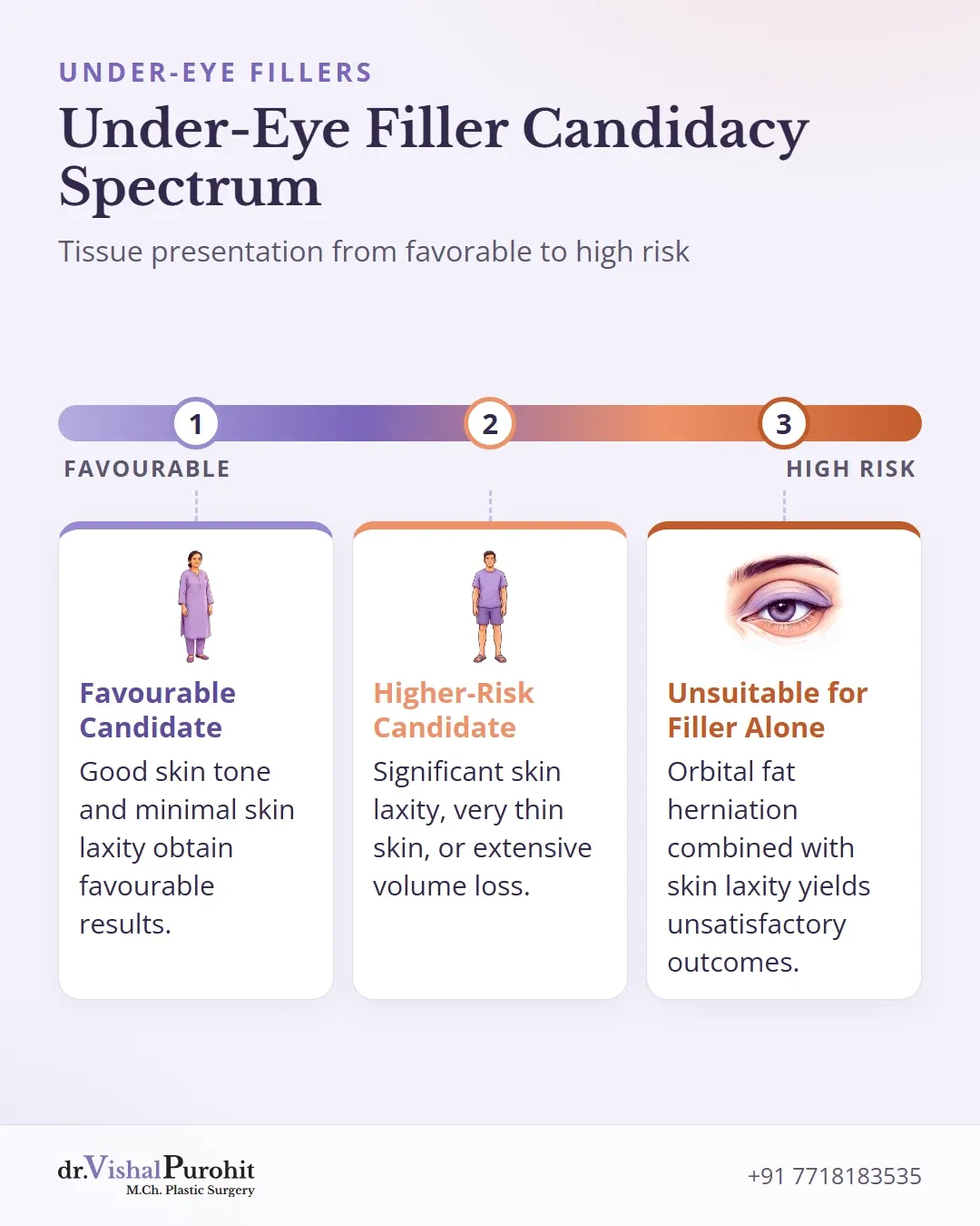
Evaluating the periorbital area requires examining tissue quality, baseline skin laxity, and the presence of puffiness or swelling1718. Patients with good skin tone and minimal skin laxity obtain favourable results, whereas those with significant skin laxity, very thin skin, or extensive volume loss represent higher-risk candidates19. Individuals with orbital fat herniation combined with skin laxity are unlikely to achieve satisfactory outcomes from injecting the tear trough alone19.
Whether an individual is suitable for under-eye filler or if alternative approaches are appropriate is decided at the consultation. Standardised photographs taken for records do not substitute for dynamic hands-on palpation during clinical evaluation2021.
Clinical Consultation for Under-Eye Rejuvenation in Jaipur
Periorbital rejuvenation requires precise clinical assessment because tear trough hollows, herniated fat pockets, and surface pigmentation require distinct approaches. In-person dynamic palpation evaluates tissue support, skin snap-back, and fluid tendencies before any procedure is planned. Learn more about candidacy for Under-Eye Fillers in Jaipur or review common questions regarding treatment suitability through the Under-Eye Fillers FAQs before booking your personal clinical evaluation.
This is educational information, not medical advice.