
Blepharoplasty is surgery on the eyelids. It can address heaviness in the upper lids that shadows your vision, and it can soften the bags and hollows that make the lower lids look tired.
But “eyelid surgery” is really two different operations. Upper blepharoplasty and lower blepharoplasty involve different tissue planes, different incisions, different risks, and different recoveries. Most of the confusion I see in consultation comes from the two being discussed as one procedure.
This article walks through the anatomy underneath both, so that when we sit down together you already know what the terms mean.
Considering eyelid surgery in Jaipur? Call +91-7718183535 to arrange a consultation, or read the full guide to blepharoplasty first.
First: droopy skin and a droopy lid are not the same problem
This distinction decides which operation you actually need, so it comes before anything else.
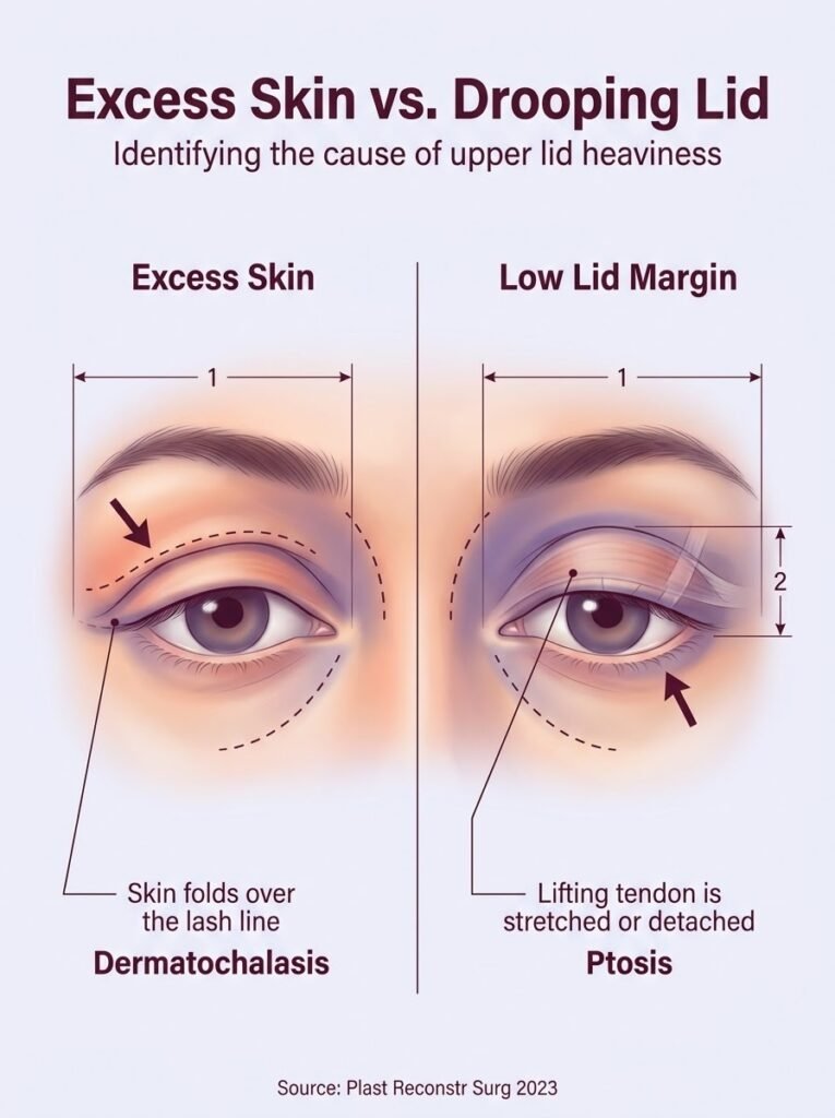
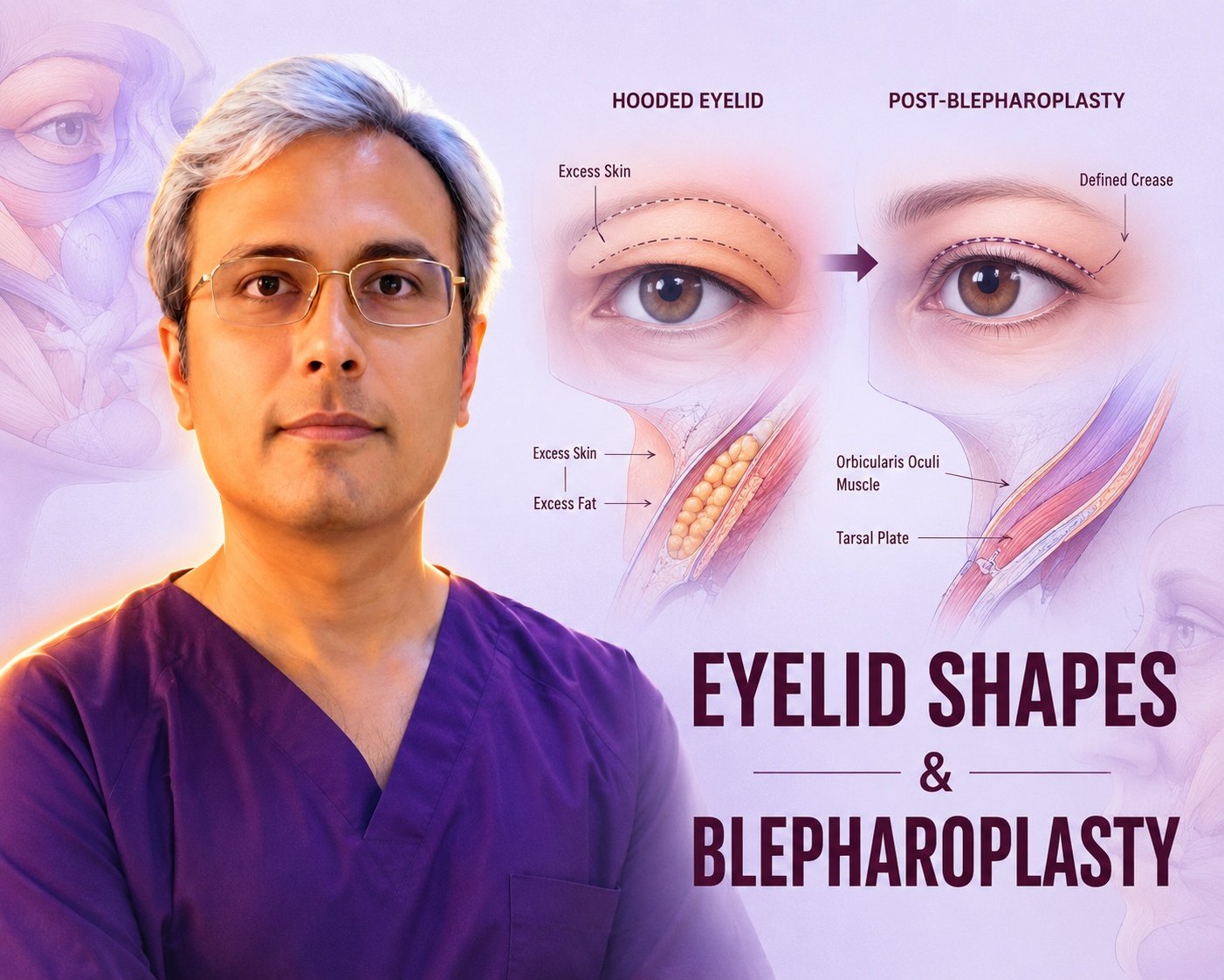
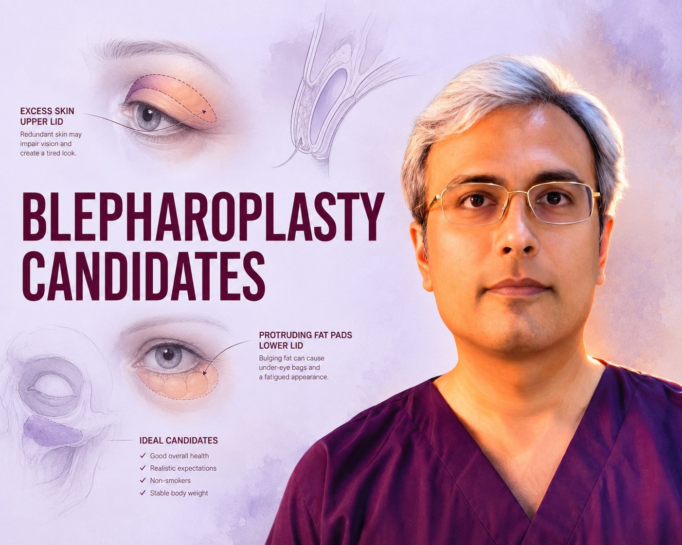
Dermatochalasis is excess upper eyelid skin. As skin loses elasticity, it folds down over the lash line, and in advanced cases it obstructs the upper part of your visual field. The lid margin itself sits where it should. Removing the redundant skin — a blepharoplasty — corrects it.
Ptosis is a low-sitting lid margin. Here the levator aponeurosis, the thin tendon that lifts the upper lid, has stretched or detached from the tarsal plate. The problem is the lifting mechanism, not the skin. Removing skin will not raise the margin; the lid needs the aponeurosis tightened, or in severe cases with poor levator function, suspension to the frontalis muscle (Cang et al., Plast Reconstr Surg 2023).
The two often coexist. A patient can have hooded skin and a low margin, and correcting only the skin leaves them looking half-treated. This is why I measure margin-to-reflex distance and levator function at the first visit rather than simply pinching skin.
Brow position is the third variable. When the brow descends, the upper lid looks heavier than it is. Operating on the lid alone in that situation can pull the brow down further. The periorbital region behaves as one connected unit, and the brow has to be assessed alongside the lid (Pascali et al., Facial Plast Surg 2023).
The structures that matter
The tarsal plate
Each eyelid contains a plate of dense fibrous tissue — not cartilage, though it feels like it — that gives the lid its shape and stiffness. The upper tarsus is taller, roughly 10 mm at its centre; the lower is about 4 mm. It is the scaffold the lash line hangs from, and the anchor point for the levator aponeurosis above and the lower lid retractors below.
Preserving the tarsus is non-negotiable. Almost every technical decision in eyelid surgery is made around it.
The canthal tendons
The tarsal plate gives the lid rigidity, but it does not hold the lid in position. That job belongs to the medial and lateral canthal tendons, which anchor each end of the lid to the orbital rim.
The lateral canthal tendon is the one that matters most in lower blepharoplasty. If it has loosened with age, the lower lid loses its snap against the globe. Operate on a lax lid without addressing that laxity and you invite the lid to drift downward afterwards — scleral show, or in the worst case ectropion. Testing canthal laxity before surgery is more predictive of a good outcome than almost anything else I check.
Where laxity is present, the tendon is reinforced or repositioned — a canthopexy or canthoplasty — as part of the same operation (Botti et al., Aesthet Surg J 2019).
The orbicularis oculi
This is the sphincter muscle that closes the eye. It runs in concentric bands around the orbit and is responsible for blinking, for the pump that moves tears toward the drainage system, and for protecting the cornea.
In lower lid surgery the muscle is often lifted, redraped and re-fixed rather than simply removed, because its tone contributes to lid support. Over-resection weakens closure and worsens dry eye.
The orbital septum and the fat pads
Behind the orbicularis lies the orbital septum, a thin fibrous sheet that holds orbital fat inside the socket. With age the septum weakens and the fat bulges forward. That bulge is what you see as under-eye bags — not extra fat, in most cases, but the same fat pushed forward.
The upper lid has two fat pads; the lower lid has three. Below the orbital rim sits a separate cushion, the suborbicularis oculi fat, which contributes to the lid–cheek transition.
The lid–cheek junction
The tear trough and the hollow beside it are largely a matter of where the orbital rim ligaments tether skin down. Understanding this is why modern lower blepharoplasty often repositions fat rather than removing it — more on that below.
Upper blepharoplasty
The incision
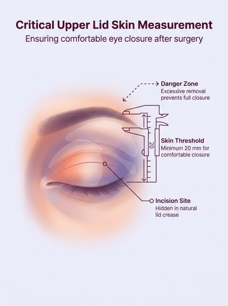
The incision sits in the natural upper lid crease. Once healed, it is hidden when the eye is open, and visible only as a fine line when the eye is closed.
The critical decision is how much skin to leave, not how much to take. A minimum of about 20 mm of skin between the brow and the lash line is needed for the lid to close comfortably. Take more than that and the patient is left unable to shut the eye fully — a difficult problem to reverse. I would rather leave a patient slightly under-corrected and revise later.
What is removed, and what is kept
Skin is the main target. A strip of orbicularis may be taken if the lid is very full. Fat is treated conservatively: the medial pad is often the only one that genuinely needs reducing, and aggressive upper lid fat removal is a common cause of the hollow, skeletonised look that reads as “operated on” rather than rested.
If the levator aponeurosis has stretched, it is repaired through the same incision at the same sitting.
Lower blepharoplasty
Two routes in
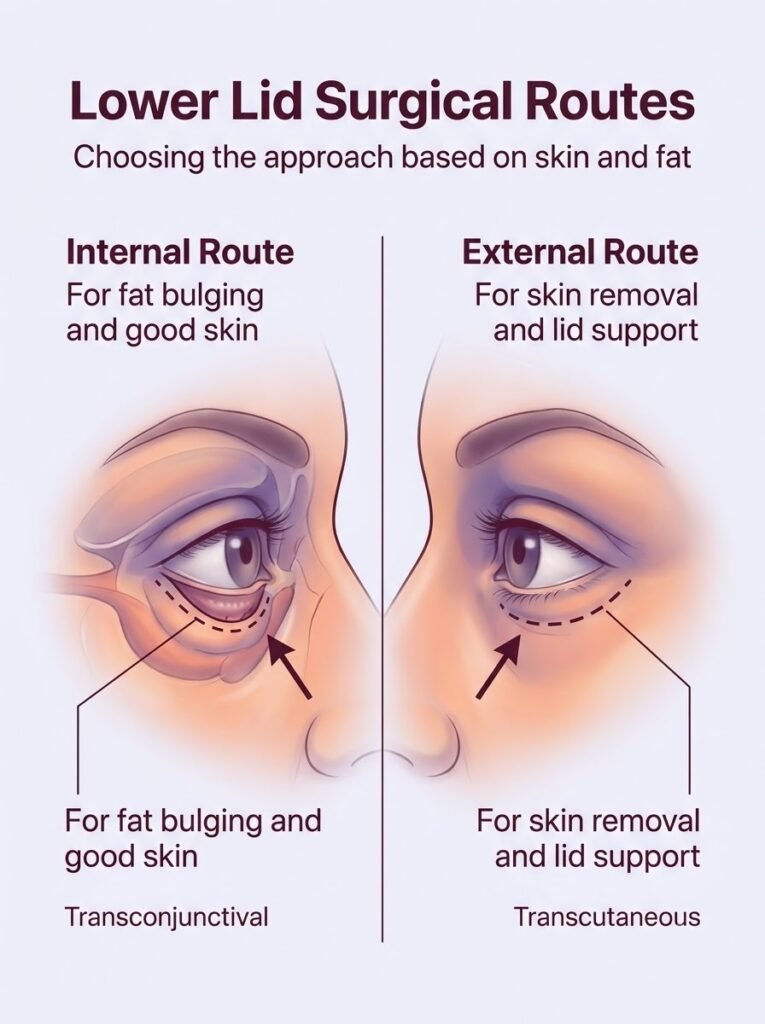
Transconjunctival — the incision is made on the inside of the lower lid. There is no external scar, and because the orbicularis and its supporting structures are left undisturbed, the risk of the lid pulling downward afterwards is lower. It suits patients whose main issue is fat bulging with good skin quality (Pacella et al., Plast Reconstr Surg 2010).
Transcutaneous — a subciliary incision just below the lash line, which allows excess skin to be removed as well as fat addressed. It is the route for patients with genuine skin redundancy, and it is usually combined with canthal support (Bhattacharjee et al., Indian J Ophthalmol 2020).
Neither is universally better. The choice follows the anatomy.
Repositioning rather than removing fat
The older approach was to excise the bulging fat. It flattened the bag but often left a hollow above the cheek, because the fat was doing volumetric work as well as causing the bulge.
The alternative is to release the tethering at the orbital rim and redrape that same fat downward over the rim, filling the tear trough with it (Hamra, Plast Reconstr Surg 1995). Combined with attention to the lid–cheek junction, this produces a smoother transition than excision alone (Rohrich et al., Plast Reconstr Surg 2011).
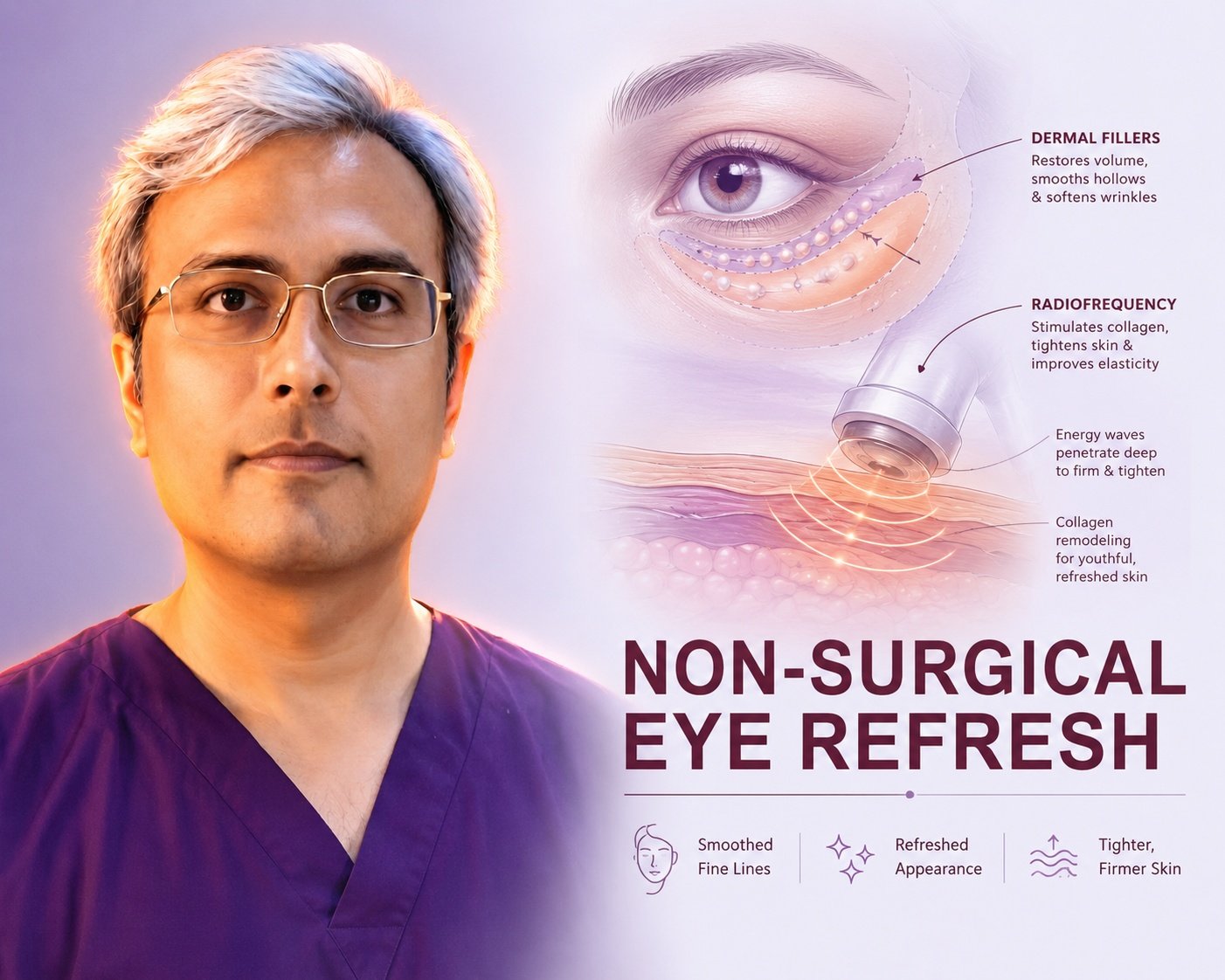
Not everyone is a candidate. Where volume loss dominates, fat grafting or filler may serve better than repositioning.
How much skin can safely come off
Very little, if the lid is not supported. Traditional teaching limits subciliary skin excision to roughly 2–4 mm precisely because over-resection is the classic route to ectropion (Rosenberg et al., Arch Facial Plast Surg 2007).
More can be taken safely, but only when the suspension system is deliberately reinforced. In a ten-year series of extended lower blepharoplasty with canthoplasty and muscle fixation, an average of 10 mm was excised with one case of ectropion across 93 patients (Fabbri et al., Aesthetic Plast Surg 2026). The lesson is not “more skin is fine” — it is that skin excision and lid support are a single decision, not two.
What I assess before agreeing to operate
This is the part of the process that most determines the result, and the part patients hear least about.
- Dry eye history and tear film. Blepharoplasty transiently worsens dry eye. A patient with pre-existing dryness, or a history of LASIK, needs that established and managed first.
- Canthal laxity. Snap-back and lid distraction testing. Laxity changes the operative plan.
- Margin-to-reflex distance and levator function. To separate ptosis from dermatochalasis, as above.
- Brow position. Assessed with the patient’s brow at rest, not raised.
- Globe position and vector. In a negative vector face — where the eye sits forward of the cheek — the lower lid is inherently less well supported and standard techniques carry more risk.
- Thyroid status. Undiagnosed thyroid eye disease changes everything about the plan.
- Bleeding risk. Anticoagulants, antiplatelets, and supplements that affect clotting.
- Photographs and asymmetry mapping. Almost everyone is asymmetric before surgery. Documenting it prevents disappointment afterwards.
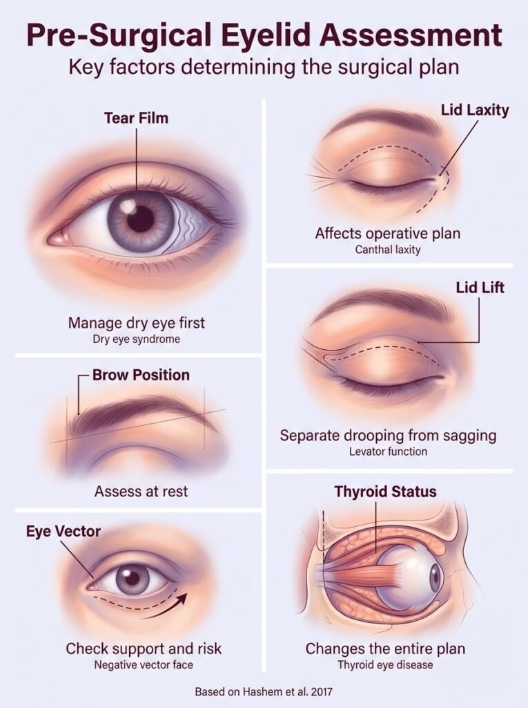
A graded, evidence-based approach to matching technique to findings has been set out in the plastic surgery literature (Hashem et al., Plast Reconstr Surg 2017), and it is broadly the framework I work within.
Risks, named honestly
Every operation carries risk. I would rather you read them here than discover them later.
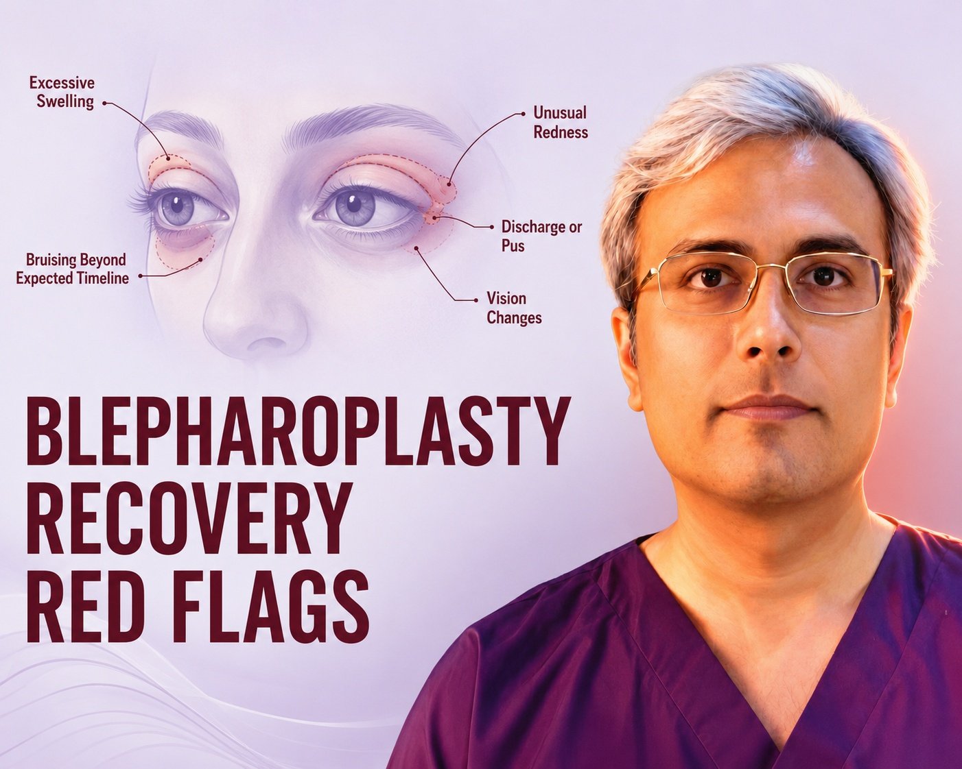
Retrobulbar haemorrhage. Rare, and the reason eyelid surgery is taken seriously. Bleeding behind the globe raises orbital pressure and can threaten vision. It presents in the first hours as increasing pain, proptosis and visual change, and it is a surgical emergency. This is why I give explicit post-operative instructions about what to watch for and how to reach me immediately.
Lower lid malposition. Scleral show, lid retraction or ectropion, usually from over-resection of skin or unrecognised canthal laxity. Often settles with massage and time; sometimes needs revision.
Chemosis. Swelling of the conjunctiva. Common enough after lower lid surgery to be worth expecting, uncomfortable, and self-limiting with treatment.
Dry eye and incomplete closure. Usually temporary. Managed with lubricants. Persistent lagophthalmos after over-aggressive skin removal is the version to avoid.
Asymmetry. Faces are asymmetric to begin with, and healing is not perfectly symmetric either.
Scarring. Upper lid scars usually settle to an inconspicuous line in the crease. Subciliary scars are generally good but take longer to fade.
Under- or over-correction, and the possibility of revision surgery.
There is a fuller discussion in my article on the common risks and safety realities of eyelid procedures.
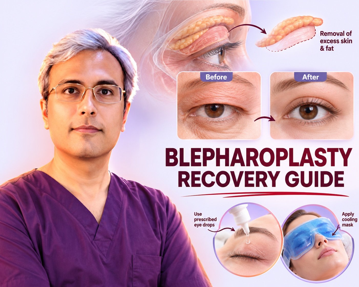
Recovery: a realistic timeline
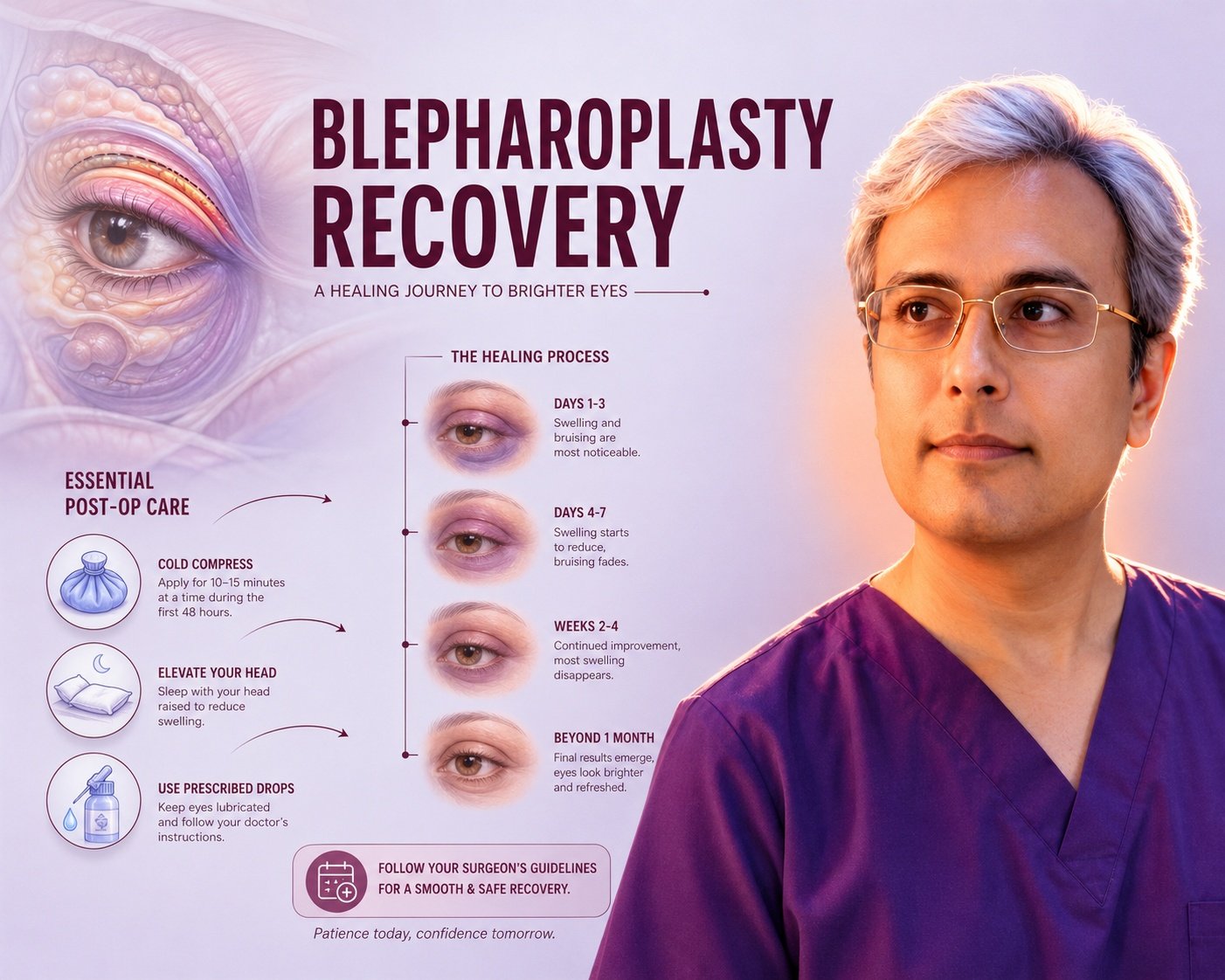
- Day 0–3. Swelling and bruising increase, peaking around 48–72 hours. Cold compresses, head elevated. Vision may be blurred from ointment. Expect to do nothing.
- Day 4–7. Sutures removed, usually in this window. Swelling starting to turn the corner.
- Day 7–14. Most patients are comfortable in public with light make-up by around two weeks. Bruising fades to yellow.
- Week 2–4. Back to desk work and light activity. Screen time may still tire the eyes. No heavy lifting or straining.
- Week 4–6. Exercise resumed gradually. Scars still pink.
- Month 3–6. Scars soften and pale. Fine residual swelling settles, particularly in the lower lids.
- Month 6–12. Final result.
Air travel is usually reasonable after about two weeks, but check with me based on your specific procedure. Contact lens wear typically resumes at two to three weeks.
Results vary considerably between individuals. Upper blepharoplasty results commonly last well over a decade; lower lid fat correction tends to be long-lasting, but neither stops the ageing process — they reset the starting point.
Aesthetic judgement
Technique keeps the eye safe. Judgement decides whether it looks like you.
The goal is not a standard eyelid. Crease height, lid show, and the degree of fullness that suits a face vary with ethnicity, gender, and personal preference, and the operation should follow the face rather than a template. I discuss this at length in defining your aesthetic goals.
If you want to talk through whether eyelid surgery is appropriate for you, cosmetic surgery in Jaipur starts with a consultation, not a procedure.
Frequently asked questions
Will the scars be visible after blepharoplasyty?
Upper eyelid incisions are placed in the natural crease, so they are hidden when your eyes are open and appear as a fine line when closed. Lower lid incisions are either inside the lid, leaving no external scar at all, or just below the lash line. Scars are pink for roughly three months and continue to fade for up to a year. Sun protection during that period makes a measurable difference.
How soon can I see and blink normally after eyelid surgery?
Blinking works from day one, but it will not feel normal immediately. Expect blurred vision from lubricating ointment for the first few days, and some tightness on full closure for one to two weeks while swelling settles. Most patients are comfortable reading and using screens for short periods by the end of the first week, and normally by weeks three to four. Persistent difficulty closing the eye is not expected and should be reported.
How long do the results of blepharoplasty last?
Upper eyelid results commonly last ten to fifteen years or more. Lower lid correction, particularly where fat has been repositioned and lid support reinforced, tends to be long-lasting. Neither halts ageing — skin continues to lose elasticity — so think of surgery as setting the clock back rather than stopping it. Individual results vary.
When will the puffiness completely go after blepharoplasty?
It depends on what is causing it. Bulging orbital fat responds well to repositioning or reduction. Puffiness caused by fluid retention, allergy, thyroid disease or lymphatic congestion may not improve with surgery at all, and malar bags in particular are notoriously resistant. Establishing the cause at consultation is what prevents a disappointing result.
Is upper eyelid surgery covered by insurance?
Where excess upper lid skin genuinely obstructs the upper visual field, the procedure may be considered functional rather than cosmetic, and some insurers will consider it. This normally requires documented visual field testing and photographs. Purely cosmetic eyelid surgery is not covered. Policies differ, so this needs checking with your specific insurer.
What anaesthesia is used in eyelid surgery?
Upper blepharoplasty alone is frequently performed under local anaesthesia with or without sedation, as a day procedure. Lower blepharoplasty, and combined upper and lower surgery, is more often done under sedation or general anaesthesia. The choice depends on the extent of surgery and your own preference and medical history.
About the author
Dr. Vishal Purohit is a plastic, cosmetic and reconstructive surgeon practising in Jaipur, Rajasthan. [Add qualifications, years in practice, hospital affiliations, society memberships and medical council registration number here — this section carries significant weight for both patients and search engines.]
Medically reviewed: 26 July 2026.
Consultation
To discuss whether eyelid surgery is appropriate for you, call +91-7718183535 or visit the clinic in Jaipur.
Disclaimer: This article is for general education and does not constitute medical advice. It cannot substitute for an in-person assessment. Surgical outcomes vary between individuals, and no specific result is guaranteed. Every procedure carries risk. Please discuss your own circumstances with a qualified plastic surgeon before making any decision.
References
- Cang ZQ, et al. Improved eyelid muscle tension balance with refined frontalis muscle flap suspension in the treatment of severe ptosis. Plast Reconstr Surg. 2023;152(5):885e–894e. doi:10.1097/PRS.0000000000010368
- Pascali M, et al. The temporal subcutaneous brow lift with orbicularis oculi muscle suspension. Facial Plast Surg. 2023;39(6):691–702. doi:10.1055/a-1953-2304
- Botti G, et al. “Dynamic canthopexy” drill hole canthal repositioning. Aesthet Surg J. 2019;39(12):1284–1294. doi:10.1093/asj/sjz077
- Pacella SJ, Nahai FR, Nahai F. Transconjunctival blepharoplasty for upper and lower eyelids. Plast Reconstr Surg. 2010;125(1):384–392. doi:10.1097/PRS.0b013e3181c2a534
- Bhattacharjee K, Ghosh S, Ugradar S, Azhdam AM. Lower eyelid blepharoplasty: an overview. Indian J Ophthalmol. 2020;68(10):2075–2083. doi:10.4103/ijo.IJO_2265_19
- Hamra ST. Arcus marginalis release and orbital fat preservation in midface rejuvenation. Plast Reconstr Surg. 1995;96(2):354–362. doi:10.1097/00006534-199508000-00014
- Rohrich RJ, Ghavami A, Mojallal A. The five-step lower blepharoplasty: blending the eyelid–cheek junction. Plast Reconstr Surg. 2011;128(3):775–783. doi:10.1097/PRS.0b013e3182121618
- Rosenberg DB, Lattman J, Shah AR. Prevention of lower eyelid malposition after blepharoplasty. Arch Facial Plast Surg. 2007;9(6):434–438. doi:10.1001/archfaci.9.6.434
- Fabbri M, et al. Extended lower blepharoplasty: how much skin can we resect? Aesthetic Plast Surg. 2026;50(1):87–95. doi:10.1007/s00266-025-05405-7
- Hashem AM, Couto RA, Waltzman JT, Drake RL, Zins JE. Evidence-based medicine: a graded approach to lower lid blepharoplasty. Plast Reconstr Surg. 2017;139(1):139e–150e. doi:10.1097/PRS.0000000000002849










 English
  Hindi
Hindi